The content of research paper

A reduction method for anterior opening displacement in thoracolumbarvertebral fractures with diffuse idiopathic skeletal hyperostosis using the skull clamp-assisted position

Hiroshi Kobayashi, Kazuyuki Watanabe, Yoshihiro Kobayashi, Kinshi Kato, Takuya Nikaido, Koji Otani, Shoji Yabuki, Shin-ichi Konno, Yoshihiro Matsumoto

Author information
  • Hiroshi Kobayashi

    Department of Orthopaedic Surgery, Fukushima Medical University School of Medicine

  • Kazuyuki Watanabe

    Department of Orthopaedic Surgery, Fukushima Medical University School of Medicine

  • Yoshihiro Kobayashi

    Department of Orthopaedic Surgery, Fukushima Medical University School of Medicine

  • Kinshi Kato

    Department of Orthopaedic Surgery, Fukushima Medical University School of Medicine

  • Takuya Nikaido

    Department of Orthopaedic Surgery, Fukushima Medical University School of Medicine

  • Koji Otani

    Department of Orthopaedic Surgery, Fukushima Medical University School of Medicine

  • Shoji Yabuki

    Department of Orthopaedic Surgery, Fukushima Medical University School of Medicine

  • Shin-ichi Konno

    Department of Orthopaedic Surgery, Fukushima Medical University School of Medicine

  • Yoshihiro Matsumoto

    Department of Orthopaedic Surgery, Fukushima Medical University School of Medicine

Introduction

Diffuse idiopathic skeletal hyperostosis (DISH) is characterized by bone hyperplasia of the skeletal system, particularly of the spine1). In this case, the spine becomes rigid, causing stress concentrations when exposed to external forces, with even minor trauma leading to a spinal cord injury2). DISH is a common indication for surgery, making patients susceptible to unstable spinal fractures due to anatomical features3).


The prevalence of DISH on computed tomography (CT) is as high as 27.2% in total, 38.7% in males, and 14.0% in females4). Moreover, the prevalence of DISH rises with increasing age5,6);therefore, this fracture is common, especially with increasing age.


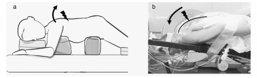
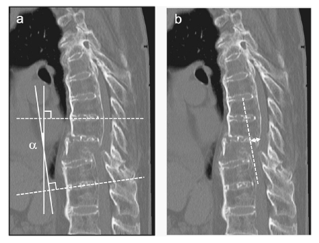
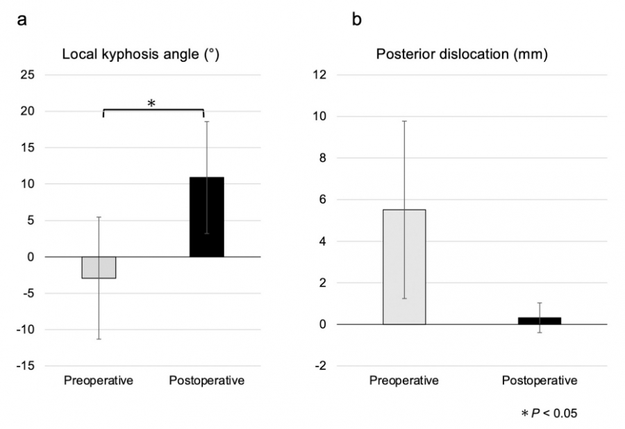
Patients with DISH often have thoracic kyphosis, resulting in loss of kyphosis or lordosis due to hyperextension injuries7). When the patient is placed in the prone position for posterior fixation with this fracture, extension forces are applied to the fracture site, exacerbating the displacement, and making the reduction challenging. If the anterior opening displacement remains after posterior fixation, the anterior column may be lacking, resulting in non-union and implant failure.


However, adding anterior fixation to avert such complications is highly invasive, increases the risk of complications, and is contraindicated in patients who may not handle the invasiveness7). Additionally, displacement of the fracture site may exacerbate neurological deficits. By performing preoperative reduction, we can achieve surgical goals with minimally invasive surgery comprising posterior fixation only. Moreover, indications for surgery could be extended, and a superior outcome could be observed. This also has the potential to prevent neurological exacerbations.


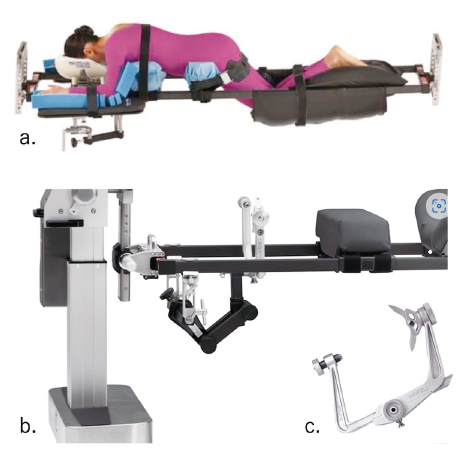
In this study, we establish a method for preventing displacement of the surgical position and approach repositioning using the skull clamp-assisted position (SAP) for thoracolumbar fractures with DISH. Additionally, we describe the technique and its precautions and report several cases treated with this method.

Material and methods

The study protocol was approved by the Ethics Committee of Fukushima Medical University (Project identification code 2022-196). For all subjects, instead of written consent, information about the management of the study was publicly available on the website, and the opportunity to refuse participation was guaranteed.


The inclusion criteria for this study were patients with unstable hyperextension fractures of the thoracolumbar spine due to low-energy trauma (type B3, according to Magerl8)) while also meeting the diagnostic criteria of DISH1), with at least three tonic segments present between the fracture’s cephalad and caudal sides.


Reduction technique


After administering general anesthesia, we placed the patient in a prone surgical position without placing their head on the operating table to avoid opening the fracture site. Instead, the head was fixed to the Modular Table System (MIZUHO, Tokyo, Japan, Fig. 1a, b), originally known as the Jackson table, using a skull clamp (MAYFIELD® Skull Clamp, Integra LifeSciences Corporation, NJ, USA. Fig. 1c). To recreate the pre-fracture alignment of the kyphosis, we immobilized the patient in the prone position, with their head lowered naturally and without strain toward the floor (Fig. 2).


To prevent increased intracranial pressure, we manipulated the tilt of the entire operating table to elevate the head. Percutaneous pedicle screws were used for fixation. The range of fixation was 3 levels above and below across the fracture in all cases. The rods were 5.5 mm titanium alloy, bent to fit the kyphosis formed by the body position, and fixed with set screws. Postoperatively, the patient was fitted with a soft brace and mobilized under the guidance of a physical therapist, who encouraged resumption of activities without specific restrictions. This approach facilitated an early discharge from the hospital. The orthosis was removed after 2-3 months.


In the CT sagittal images taken preoperatively and one week postoperatively, the difference in local kyphosis angle (°, Fig. 3a) and posterior dislocation of the cranial vertebrae compared to the fractured vertebra (mm, Fig. 3b), and complications up to one year after surgery (namely neuropathy, implant failure, and reoperation due to infection) were also evaluated. The local kyphosis angle was defined as the angle formed between the upper endplate of the vertebra above the fracture and the lower endplate of the vertebra below the fracture, as evidenced in the CT sagittal image.


Fig. 1. Operating table used for skull clamp-assisted position and skull clamp

(a) Modular Table System (MIZUHO, Tokyo, Japan), originally known as the Jackson table. (b) Attachment to connect Modular Table System and skull clamp (MIZUHO, Tokyo, Japan). (c) Skull clamp (MAYFIELD® Skull Clamp, Integra LifeSciences Corporation, NJ, USA).


Fig. 2. Surgical position

(a) Conventional surgical position. The patient is positioned using a four-point frame. The arrows indicate the forces exerted by the head, and thoracic supports to dislocate the head side of the fracture dorsally and open the fracture anteriorly. (b) Surgical position using the skull clamp and Jackson table. The patient is positioned to reproduce the preoperative alignment of the thoracic spine by attaching a skull clamp and positioning the head down (arrow). The cranial side of the fracture is maintained in its natural pre-fracture alignment, preventing the progression of displacement. The entire bed is tilted to elevate the head side to prevent increased intracranial pressure (arrowhead).


Fig. 3. Measurements of radiographic image parameters

(a) Local kyphosis angle: α (degrees). (b) Posterior displacement of the cranial vertebra: double arrow (mm).


Statistical analyses


The JMP®, Pro version 16.0.0 (SAS Institute Inc., Cary, NC, USA) was used for statistical analysis. Differences in the local kyphoscoliosis angle and posterior dislocation of the cranial vertebrae were compared using Student’s t test;a P < .05 was considered statistically significant.

Results

The five cases are presented in Table 1. The series comprised 3 males and 2 females, with a mean age of 81.2 years. The fractured vertebral levels ranged from T6 to L1, and all fractures were classified as AO type B3. Two cases presented with nerve palsy, and in one case, decompression was performed in addition to fixation.


The mean difference in local kyphosis angle before surgery was -2.9 (±8.4)°, whereas that after surgery was 10.9 (±7.7)°, showing significant improvement (P = .0052) (Fig. 4a). The mean difference in posterior displacement before surgery was 5.5 (±4.3) mm, whereas after surgery was 0.3 (±0.7) mm, showing a trend toward improvement. However, the difference was not significant (P = .063) (Fig. 4b). No complications, such as neurological sequelae, implant failure, or surgical site infection, were observed for up to one year postoperatively.


Table 1. Demographic, Surgical, and Radiolgraphical Characteristics of the 5 Cases.

M, male;F, female


Fig. 4. Changes in radiographic image parameters before and after surgery

The local kyphosis angle was significantly improved postoperatively by creating a surgical position using the skull clump.


Case presentation (Case 4 in Table 1, Fig. 5)


The case involved an 87-year-old female patient who sustained a back injury due to a fall. Nine days after the fall, she was admitted to a prior hospital with a diagnosis of T9 compression fracture, where conservative treatment was initially considered.


One month post-fall, she developed urinary retention and paralysis in both lower limbs, grade 0-3 on the Manual Muscle Testing (MMT), which led to her referral from the previous hospital. The diagnosis of a T9 vertebral fracture (AO type B3) with delayed onset paralysis associated with DISH necessitated emergency surgery.


The surgical procedure involved posterior fixation using percutaneous pedicle screws from T6 to T12. The kyphosis was 4.0˚ preoperatively and improved to 23.6˚ postoperatively. The paralysis improved to a grade above 3 on the MMT. At 6 months postoperatively, the patient was able to walk with a pickup walker. Bone union was achieved 10 months post-surgery, and no complications were observed at the one-year follow-up.


Fig. 5. Representative case (Case 4 in Table 1)

(a) The preoperative sagittal computed tomography (CT) image in the supine position shows the anterior opening of the vertebral body of T9 (arrow), the posterior displacement of the cranial side, and the vertebral body’s reduced height. (b) Postoperative CT sagittal image in the supine position showing the anterior opening of the vertebral body of T9, the cephalad posterior displacement, and the height of the vertebral body have been realigned (arrowhead).

Discussion

There are several problems with surgery for thoracolumbar fracture with DISH. First, this fracture type is highly unstable, with previous reports recommending extensive fixation9). Second, the bone is osteoporotic, and the fixation of the screw is not strong. Consequently, there have been reports of innovations regarding screw insertion, such as developing transdiscal screws10), cement augmentation for percutaneous pedicle screws11), and the efficacy of teriparatide12), which are considered to improve treatment outcomes. Third, a displacement that opens the anterior vertebral body in DISH vertebral fractures is often encountered due to an extension-type injury mechanism. In DISH, kyphosis often tends to be more potent13), and bruising of the back may lead to an extension fracture, opening the fracture site of the anterior vertebral body. Various methods of repair have been reported for anteriorly opened displacements. For example, Reinhold et al. reported surgery in the sitting position14). Additionally, Lindtner reported a fixation technique using an less rigid rod, 5.5 mm pure titanium, and a percutaneous pedicle screw without aggressive fracture reduction. This method allowed for postoperative mobilization of the fracture and achieved successful results15). Ikuma et al. reported preventing anterior widening through percutaneous pedicle screw puncture in the lateral recumbent position16). However, these methods require sufficient preparation and experience. Therefore, there has been no definitive solution for managing this condition.


We have devised a surgical position to manage open anterior displacement. Conventionally, the cranial side of the fracture is compressed by the head frame and a four-point support with contact at the nipple level, resulting in an extension of the thoracolumbar spine and worsening the displacement of the fracture (Fig. 2a). If ankylosis is additionally observed from the cervical to thoracic spine, the force to open the anterior vertebral body must be even stronger. In this novel SAP method, a skull clamp was used to lower the head, which may have allowed a reduction of the displacement of the extension fracture (Fig. 2b, 5b).


When performing this technique, it is essential to note that many patients with DISH are large in stature, and the C-arm radiography is challenging to see because of the kyphosis position with a narrow working space, rendering it challenging to operate. One possible solution is to use an operating table with space below the operating table, such as the Jackson table, to create sufficient working space. Navigation systems are also beneficial in eliminating the need for a C-arm, solving the working space problem, and facilitating orientation for screw insertion. Another problem is the likelihood of increased intracranial pressure, as prone positioning has been reported to increase cerebral pressure17). Using the skull clamp to recreate the original kyphosis may further increase intracranial pressure. As the head is lowered due to kyphosis, it may be necessary to tilt the operating table to elevate the lowered head after the positioning to avoid intracranial pressure buildup as much as possible.


This method has several advantages. First, if the patient can achieve a reduction in the prone position and retainment with posterior fixation alone, it is expected to be significantly less invasive. This would reduce complications and expand the range of indications to include patients previously considered intolerant to surgery. Second, there could be a neurologic benefit by reduction. Previously, it was noted that 14.5% of patients operated on for vertebral fractures with DISH reported worsening neurologic status three months postoperatively2). This reduction technique may contribute to the amelioration of nerve compression and improve postoperative outcomes.


Finally, physical and mental workload has been considered essential in evaluating new surgical techniques18). Spine surgeons routinely use this technique in cervical spine surgery, which is considered superior. This study has some limitations. First, the number of cases is small. Therefore, we could not examine factors related to the restoration, such as the fracture site, the effect of posterior element injury, or the effect of the ankylosis site. Second, since this study examined short-term results, long-term follow-up is needed. Finally, the objective of proper alignment remains to be validated. Whether kyphotic alignment before injury is optimal remains a matter of discussion.


In conclusion, SAP effectively prevented anterior open thoracolumbar fractures in DISH from being aggravated by the surgical position and repositioned displacements closer to their original alignment. This technique should be considered an easy, safe, and effective tool for treating this trauma.

Acknowledgment

We thank Dr. Masataka Nakamura for his assistance in submitting this manuscript.

Conflict of Interest

The authors declare no relevant conflicts of interest.