Patients
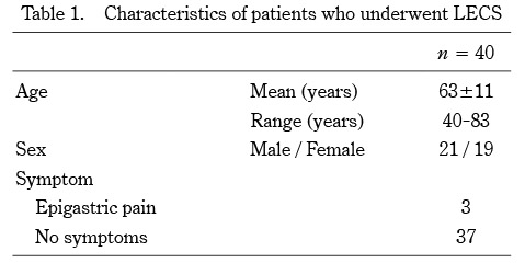
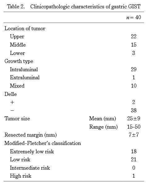
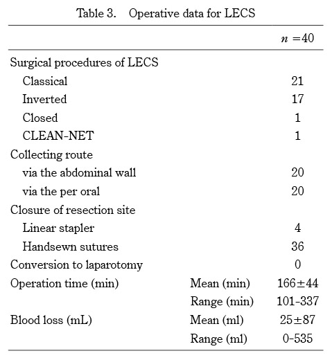
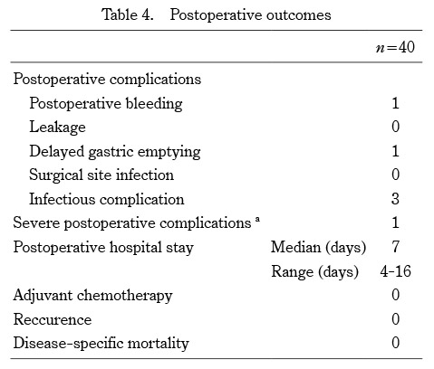
Between September 2012 and December 2020, a total of 40 patients with gastric GIST underwent LECS at the Department of Gastrointestinal Tract Surgery, Fukushima Medical University Hospital (Fukushima, Japan). The patients’ hospital records were retrospectively analyzed to assess patient background, perioperative and postoperative outcomes, as well as the tumor clinicopathological characteristics. The following surgical data were included in the analyses:the collection route of the resected tumor; the resection site closure method; the operation time; and the estimated blood loss. The following postoperative data were collected; modified-Fletcher’s classification, tumor size, postoperative complications (bleeding, leakage, delayed gastric emptying, infection including surgical site infection), postoperative hospital stay length, tumor recurrence, and mortality.
The preoperative workup for all patients included reviews of the medical history, standard blood tests, an upper gastrointestinal endoscopy with endoscopic ultrasonography examination, and computed tomography. All patients had previously undergone endoscopic ultrasonography-guided fine-needle aspiration and were histologically diagnosed as having a gastric GIST by immunohistochemistry.
The surgical treatment algorithm for GIST in our hospital is shown in Figure 1. LECS for gastric GIST was not indicated for patients with a previous history of gastrectomy or multiple laparotomies. LECS was indicated for gastric GISTs of ≤ 50 mm in diameter in the preoperative diagnosis and for tumors with intraluminal or intramural growth, excluding those with typical extraluminal growth. Since 2017, all patients with intraluminal or intramural tumors in our hospital have undergone inverted LECS according to our treatment protocol, whereas CLEAN-NET or closed LECS was performed on the tumors with delle, depending on the tumor size.
Patients underwent postoperative CT scans and endoscopies at 6 months, 1 year, and then annually thereafter to diagnose and monitor any local or distant recurrence.
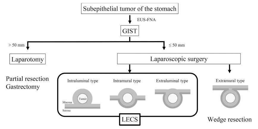
Fig. 1. Surgical treatment algorithm for gastric GIST in our hospital
Surgical procedure of classical LECS
The previous endoscopic and laparoscopic approaches in the classical LECS procedure were as follows. The patients are placed in the lithotomy position under general anesthesia and the operating room laid out as shown in Figure 2. A laparoscopic camera port is inserted into the umbilicus with the open technique. Four additional ports (one 12-mm port and three 5-mm ports) are inserted into the upper right, lower right (12-mm port), upper left and lower left quadrants under a 10 mmHg pneumoperitoneum by carbon dioxide gas. The tumor is completely resected endoscopically using an endoscopic submucosal dissection technique and full layer dissection of the gastric wall. The stomach wall defect is sutured using a linear stapler or a laparoscopic hand-sewn technique with a self-anchoring barbed suture. This combined endoscopic and laparoscopic approach enables us to resect the tumor lesion with as narrow a margin as possible. Resected specimens sized ≤ 30 mm in diameter are put in collection bags and retrieved per orally, while specimens sized > 30 mm are retrieved via the laparoscopic camera port site at the umbilicus.
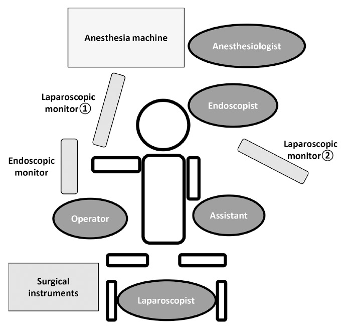
Fig. 2. Setup for LECS
Surgical procedure of inverted LECS
The routine endoscopic and laparoscopic approaches in the inverted LECS procedure are as follows. After identification of the tumor under laparoscopy and endoscopy, the gastric wall around the tumor is lifted circumferentially by three or four stitches. Each of the stitches is appropriately pulled out of the abdominal port site like a crown4). Then, the tumor is completely resected endoscopically using an endoscopic submucosal dissection technique and full layer dissection of the gastric wall. The stomach wall defect is sutured using a laparoscopic hand-sewn technique with a self-anchoring barbed suture.
Surgical procedures of CLEAN-NET and closed LECS
Inoue et al. and Kikuchi et al. developed novel non-exposure techniques, termed ‘CLEAN-NET’ and ‘closed LECS’, respectively5, 6). CLEAN-NET is a procedure to harvest the tumor into the abdominal cavity, while closed LECS is a procedure to harvest the tumor into the gastric tube cavity. We perform these surgical procedures based on the description in the original articles. Briefly describing CLEAN-NET, the surgery is performed by incising the serosal muscular layer around the tumor in the abdominal cavity without destroying the mucosa. Once the serosa of the tumor is dissected circumferentially, the full layer is lifted, and the mucosa is stretched. Then, the mucosa is cut with a stapling device, and a full-layer resection of the specimen is achieved. Both procedures enable resection without perforation of the stomach lumen and the abdominal cavity. In contrast, in the closed LECS, a circumferential incision is made endoscopically around the tumor using the endoscopic submucosal dissection technique, and then the serosal muscle layer corresponding to the submucosal dissection line is sutured in a straight line laparoscopically. Consequently, the gastric wall containing the tumor protrudes into the gastric cavity. Finally, an endoscopic serosal muscle layer incision is performed along the submucosal dissection line, and the entire gastric wall is dissected. The gastric wall defect caused by tumor resection is endoscopically closed with clips, and the resected lesion is orally retrieved. CLEAN-NET is indicated for tumors of > 30 mm in diameter with delle. On the other hand, closed LECS is indicated for tumors of ≤ 30 mm in diameter with delle, because the resected specimen needs to be retrieved orally.
Ethics Statement
This prospective observational study was approved by the Fukushima Medical University Certified Review Board (no. 3475).
Human rights statement and informed consent
All procedures were performed in accordance with the ethical standards of the relevant committees on human experimentation (institutional and national) and with the Helsinki Declaration of 1964, as well as its later versions. Informed consent or a substitute for it was obtained from all patients regarding their participation in the study.