The cintent of reseach paper

Association between the presence of bacteria in prostate tissue and histopathology in biopsies from men not complaining of lower urinary tract symptoms

Ruriko Honda-Takinami, Junya Hata, Kanako Matsuoka, Seiji Hoshi, Tomoyuki Koguchi, Yuichi Sato, Hidenori Akaihata, Masao Kataoka, Soichiro Ogawa, Kyoko Nishiyama, Tatsuo Suzutani, Yoshiyuki Kojima

Author information
  • Ruriko Honda-Takinami

    Department of Urology, Fukushima Medical University School of Medicine

  • Junya Hata

    Department of Urology, Fukushima Medical University School of Medicine

  • Kanako Matsuoka

    Department of Urology, Fukushima Medical University School of Medicine

  • Seiji Hoshi

    Department of Urology, Fukushima Medical University School of Medicine

  • Tomoyuki Koguchi

    Department of Urology, Fukushima Medical University School of Medicine

  • Yuichi Sato

    Department of Urology, Fukushima Medical University School of Medicine

  • Hidenori Akaihata

    Department of Urology, Fukushima Medical University School of Medicine

  • Masao Kataoka

    Department of Urology, Fukushima Medical University School of Medicine

  • Soichiro Ogawa

    Department of Urology, Fukushima Medical University School of Medicine

  • Kyoko Nishiyama

    Department of Microbiology, Fukushima Medical University School of Medicine

  • Tatsuo Suzutani

    Department of Microbiology, Fukushima Medical University School of Medicine

  • Yoshiyuki Kojima

    Department of Urology, Fukushima Medical University School of Medicine

Introduction

The detailed pathogenesis of benign prostatic hyperplasia (BPH) remains unclear. A previous study reported that patients with pathologically diagnosed BPH had viable bacteria in 55.5% (20 of 36) of patient tissues, with 16SrRNA sequencing detecting mainly Staphylococcus (22%), E. coli (11%), and Micrococcus spp. (8%)1). It has been also reported that E. coli was detected in the prostate tissue of 3% of patients who underwent transurethral resection of the prostate (TURP)2). However, it is not clear how the presence of bacteria in prostatic tissue affects the development of BPH.


On the other hand, there have been reports of histological evaluation and BPH assessment in experiments using purposefully infected mice; Ruetten and Wong reported that transurethral injection of E. coli, a causative agent of urinary tract infections, induced prostatic inflammation with epithelial and stromal hyperplasia and tissue fibrosis, indicating that acute bacterial infection may contribute to prostatic enlargement. They further showed that prostatitis is triggered by urinary tract infection, characterized by the fibrotic response of the prostate to inflammation, and related the amount of collagen deposition to the severity of inflammation3,4). Although the above studies suggest an association between urinary bacterial infection in the prostate and the development of BPH in mice, such reports in human subjects are lacking. In the present study, we evaluated samples from prostate biopsies in patients not reporting any obvious lower urinary tract symptom (LUTS), and assessed the presence or absence of bacteria in the prostate samples by DNA sequencing, in order to see if bacterial infection might affect histopathologic findings in the prostate.

Materials and methods

Patient recruitment


The study considered 101 patients with suspected prostate cancer who underwent transperineal prostate biopsy at Fukushima Medical University Hospital, Fukushima, Japan, between May 2019 and July 2020. Patients with prostate-specific antigen (PSA) > 4.0 ng/mL who provided written informed consent were enrolled to this study. Exclusion criteria were patients with urinary tract infection (UTI) detected in urine analysis, urothelial carcinoma detected by urinary cytology and/or magnetic resonance imaging (MRI), and history of oral antibiotics within 3 months before prostate biopsy. Ultimately, 97 patients were included in this study, none of whom had any obvious LUTS. The study protocols were approved by the ethics committee at our institution (clinical trial registration number 3671).


Sample collection and processing


To investigate the presence of bacteria in the prostate, prostate tissues were collected by ultrasound-guided transperineal prostate biopsy in an operating room under a clean field. After disinfecting the perineum, the first biopsy tissue was obtained as a study sample from the transitional zone of the right or left lobe. Biopsy samples were frozen in liquid nitrogen immediately after biopsy, and stored at −80°C. To investigate pathological findings in the prostate, 12 prostate biopsy tissues were also collected and fixed in 10% formalin.


Detection and identification of bacterial species


The presence and phylotypic profiles of bacteria found in the prostate samples were investigated. DNA extraction was performed on frozen prostate samples using QIAamp® DNA Mini Kit (QIAGEN, Tokyo, Japan ) according to the manufacturer’s instructions. Using the DNA samples, the 16S rDNA fragments were amplified by polymerase chain reaction (PCR) as follows. For detection of common bacteria, we used two universal primer sets with different specificities, 519B (8UA and 519B) and 1492R (8UA and 1492R), to enhance sensitivity in order to decrease the likelihood of false-negative results. The primer sequences and PCR conditions with KOD FX® (TOYOBO Co. Ltd., Osaka, Japan) are summerized in Table 1. Moreover, chlamydia detection was carried out using more specialized primers with nested PCR using the EmeraldAmp>® MAX PCR kit (Takara Biomedical Inc., Shiga, Japan). The PCR products were ligated with pGEM-T vector with a T-cloning method and then transfected into E. coli. After overnight culture, approximatory 10 colonies from one sample were picked and nucleotide sequences of cloned 16SrDNA fragments were determined. Primers 8UA, 519B, and 1492R were used to sequence general bacteria, and CTM1 and CTM8 or CTM4 and CTM7 for sequencing of Chlamydia trachomatis (Table 1). Sequencing was performed on an Applied Biosystems 3130xl Genetic Analyzer (Thermo Fisher Scientific, Inc., MA, USA). Bacterial species in the samples were identified by searching the sequence data for more than 98% homology in GenBank. According to the presence or absence of bacteria in the prostate tissues, the patients were divided into two groups; bacteria-present and bacteria–absent groups.


Table 1. Primers and PCR protocols used to identify bacteria in the prostate

To detect bacteria in prostate samples, 16S rDNA fragments were amplified by polymerase chain reaction (PCR). Two universal primer sets, 519B (8UA and 519B) and 1492R (8UA and 1492R), were used to detect common bacteria. For detection of chlamydia, more specialized primers were used in nested PCR. Primers and their respective PCR protocols are shown.


Histopathological analysis


Prostate biopsy tissues were fixed in 10% buffered formalin and embedded in paraffin. Tissue sections of 3 µm were prepared, stained with hematoxylin-eosin (HE), and examined at 200× magnification. We compared the pathological findings as described below between the bacteria-present and -absent groups.


1. Malignancy in the prostate tissue


Two pathologists analyzed 12 biopsy tissues from each patient in order to identify the presence or absence of prostate cancer, which was scored according to ISUP (International Society of Urological Pathology) guidelines5).


2. Stroma-to-gland ratio


The stroma-to-gland ratio in the prostate tissues of patients without evidence of malignancy was also investigated6). To assess the stromal and gladular components in prostate tissues, the epithelium and the stroma were measured in three randomly selected microscopic fields of each tissue section. The average stroma-to-gland ratio was calculated in each case.


Assessment of lower urinary tract symptoms and function


Questionnaires to evaluate LUTS were obtained the day before prostate biopsy. LUTS were measured using the International Prostate Symptom Score (IPSS) and Overactive Bladder Symptom Score (OABSS). Lower urinary tract function was also assessed using the maximum flow rate (MFR) and the voided volume (VV) in uroflowmetry (UFM), and post-voided residual urine volume (PVR) was determined by ultrasonography the day before prostate biopsy. We evaluated the differences in these parameters between the bacteria-present and -absent groups.


Statistical analysis


All values are presented as mean ± SD. The Mann-Whitney U test was used to test for significant differences between the groups. A P-value < 0.05 was chosen as the threshold for significance. All statistical analyses were carried using SPSS version 24 (IBM, Armonk, NY, USA).

Results

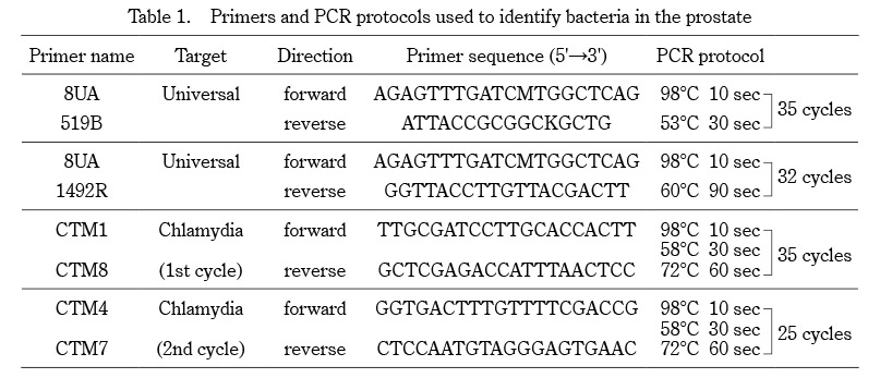
Prostate biopsies were taken from 97 patients. The presence or absence of bacteria was assessed by sequencing as described. The identification of 16S sequences indicated the presence of bacteria in the prostates of 7 patients (7.2%). Five genera of bacteria were identified by their 16S sequences; Streptococcus mitis (1/97, 1.0%), Staphylococcus haemolyticus (1/97, 1.0%), Chlamydia trachomatis (3/97, 3.1%), Cutibacterium acnes (1/97, 1.0%), and Acidovorax sp (1/97, 1.0%) (Figure 1). One bacterial species was detected in each patient, and no patient had more than one bacterial species.


To clarify associations between patient background and the presence of bacteria in prostate tissue, we compared age, height, body weight, presence of comorbidities such as hypertension and diabetes, PSA, and prostate volume between bacteria-present and -absent groups. There were no statistically significant differences in patient characteristics between the two groups (Table 2).


Differences in histological findings between the bacteria-present and -absent groups were evaluated. Of the patients with bacteria in the prostate, only one had histological evidence of malignancy, with no significant difference in the proportion of patients with prostate cancer between the two groups (p = 0.10) (Table 3). Moreover, there was no significant difference in ISUP Grade between the two groups (p = 0.12).


Next, we compared the stroma-to-gland ratio in prostate biopsies without malignant findings (n = 54) between the two groups. The stroma-to-gland ratio in the bacteria-present group was lower than that in the bacteria–absent group, suggesting that glandular components were predominant in bacteria-present group (p < 0.01) (Figure 2). In addition, histological evaluation revealed that prostate glands in the bacteria-absent group had a uniform monolayer arrangement. On the other hand, prostate glands in the bacteria-present group had ductal structures longer in shape, with irregularities in their nuclear arrangement, suggesting that glandular epithelial hyperplasia had developed in the bacteria-present group.


We also evaluated the association between the presence of bacteria in prostate tissues and LUTS. There were no significant differences in LUTS evaluated by IPSS, QOL score, or OABSS between the two groups. In addition, MFR, VV, and PVR were examined to determine the relationship between the presence of bacteria in prostate tissues and lower urinary tract function. Again, no significant differences emerged between the two groups. (MFR, p = 0.630; VV, p = 0.710; PVR, p = 0.730) (Table 4).


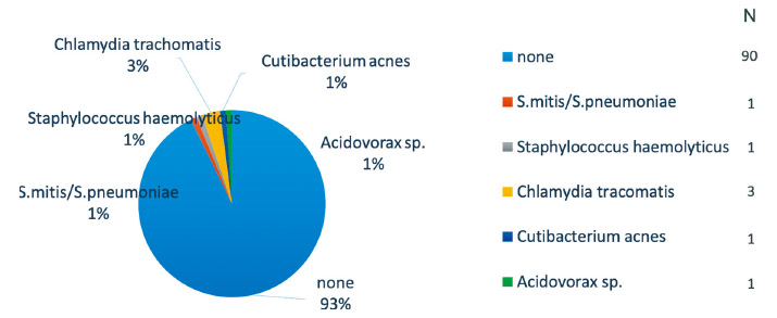
Fig. 1. Bacteria in prostate tissues

Bacteria were present in the prostates of seven patients (7.2%). Chlamydia trachomatis was the most common (3/97, 3.1%), followed by Streptcoccus mitis (1/97, 1.0%), Staphylococcus haemolyticus (1/97, 1.0%), Chlamydia trachomatis, Cutibacterium acnes (1/97, 1.0%), and Acidovorax sp. (1/97, 1.0%).


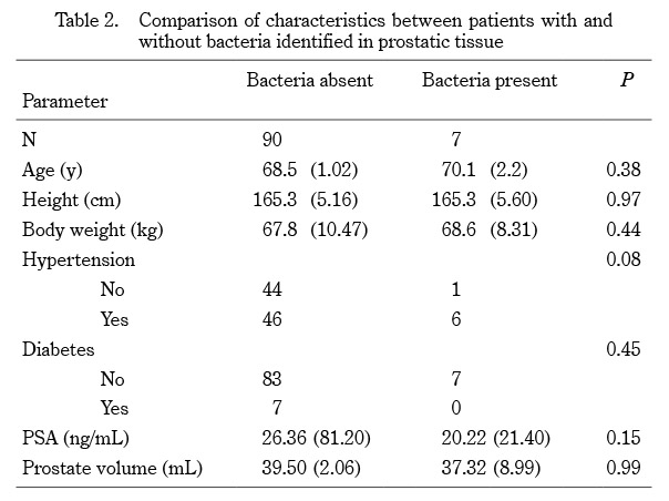
Table 2. Comparison of characteristics between patients with and without bacteria identified in prostatic tissue

Mean (SD: standard deviation)

PSA, prostate-specific antigen. Statistical significance threshold, P < 0.05.


Table 3. Association between the presence of bacteria in prostate tissue and prostate cancer

ISUP, International Society of Urological Pathology.

Statistical significance threshold, P < 0.05.


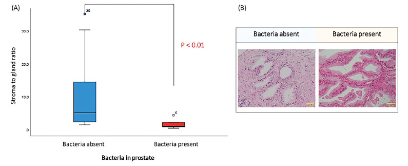
Fig. 2. Stroma-to-gland ratio and the presence or absence of bacteria in prostate tissues

The stroma-to-gland ratio in prostate tissues without malignant findings (n = 54) was compared in cases without bacteria (n = 48) and with bacteria (n = 6). The stroma-to-gland ratio in cases with bacteria was significantly lower than that in cases without bacteria. This suggests an association with an increase in glandular duct structure (A). Representative cases are shown in (B) (200×).


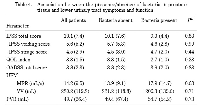
Table 4. Association between the presence/absence of bacteria in prostate tissue and lower urinary tract symptoms and function

IPSS, International Prostate Symptom Score; QOL, Quality of Life; OABSS, Overactive Bladder Symptom Score; UFM, Uroflowmetry; MFR, maximum flow rate; VV, voided volume; PVR, post-voided residual urine volume.

Mean (SD: standard deviation). Statistical significance threshold, P < 0.05.

*Bacteria-absent group vs Bacteria-present group

Discussion

We investigated the presence of bacteria in human prostate tissue from patients without obvious LUTS, and identified several bacterial species present in Japanese prostates. We showed that bacteria were present in 7.2% of prostates from patients without complaints or other evidence of LUTS.


In the present study, five bacterial species were detected:Streptococcus mitis, Staphylococcus hemolyticus, Chlamydia trachomatis, Cutibacterium acnes, and Acidovorax sp. Streptococcus sp. is known as an abscess-forming bacterium, prone to forming foci of infection such as liver and brain abscesses7). In particular, Streptococcus mitis is known as an opportunistic infectious organism. Staphylococcus sp. is known as an opportunistic infectious organism causing pneumonia and sepsis8). Chlamydia trachomatis is a causative agent of sexually transmitted disease manifesting as male urethritis, epididymitis, and orchitis9). Cutibacterium acnes is known to cause infective endocarditis, septicemia, and sarcoidosis10). Acidovorax sp. is a known phytopathogenic bacterium that parasitizes plants belonging to the Poaceae family11). Cutibacterium acnes and Acidovorax sp. are known to cause prostatic infections. However, neither of these bacteria has been reported to be endemic to the prostate. In the present study, the five species of bacteria mentioned above were present in prostate tissues from the patients without LUTS and UTI, which is a new finding in this study. Since these are not indigenous bacterial flora in the prostate, we suspected some association with conditions such as prostate cancer and BPH and, therefore, the possibility that these bacteria could cause histological changes in prostate tissue.


Previous studies have focused on associations between bacteria and the incidence or progression of prostate cancer, and have reported that bacteria are present in 19.6-95% of prostate tissues12-15). Several studies also reported that Enterobacteriaceae, E. coli, and other species of intestinal bacteria were detected in prostate tissues from surgical procedures in patients with prostate cancer, and it is thought that the preoperative insertion of catheters may have been a contributing factor16, 17). In the present study, very few bacteria were detected in just 1 of 43 patients (2.3%) diagnosed with prostate cancer by prostate biopsy. Reasons why no association emerged between the presence of bacteria in prostate tissues and the development of prostate cancer or ISUP grading might be because of the small number of bacteria-positive cases and the small volume of prostate tissue to examine for the presence of bacteria.


A previous report showed that bacterial invasion of the prostate gland is associated with the development of BPH. In an animal study, transurethral administration of E. coli induced inflammation with inflammatory cytokines, which promoted fibrosis of the prostate gland3). However, similar reports examining the effects of bacteria in human prostate tissues are lacking. BPH is pathologically defined as enlargement of both glandular components and stromal components. Kato et al. reported that glandular duct enlargement is one of the early manifestations of BPH based on age and frequency of glandular and stromal growth. Pietilä et al. also reported that mitotic activity of the glandular epithelium and glandular epithelial hyperplasia had a positive correlation with prostate weight18). These findings suggest that glandular epithelial hyperplasia might precede stromal hyperplasia during the pathogenesis of BPH. In the present study, the bacteria-present group had abundant glandular components and a lower stroma-to-gland ratio than the bacteria-absent group. Furthermore, the findings of glandular epithelial hyperplasia, including ductal structures of longer shape with irregularities in nuclear arrangement, were more abundantly observed in the bacteria-present group compared to the bacteria-absent group. Previous studies of spontaneously hypertensive rats have reported not only ventral prostate hyperplasia, but also, histological features of glandular epithelial hyperplasia in rat prostate tissues19). These results suggest that the presence of bacteria might stimulate relatively abundant increases in glandular components. In other words, the presence of bacteria in the prostate tissues might contribute to pathological changes in the early stages of benign prostate enlargement before the emergence of LUTS. Since the early stages of prostate growth were analyzed in our study, there might be no differentiation of prostate volume between the two groups.


Furthermore, the possibility that the presence of bacteria in prostate tissues could affect LUTS by prostatic inflammation was examined. Our results showed that there was no significant association between the presence of bacteria in the prostate tissues and LUTS. The patients included in this study did not have obvious clinical symptoms. Additionally, they had low IPSS, QOL index, and OABSS, and no obvious lower urinary dysfunction (LUTD). These results suggest that the presence of bacteria in the prostate does not always induce LUTS and LUTD.


Limitations of this study should be considered. The study was conducted at a single institution with a patient sample size less than 100. It also included patients with high PSA levels, which does not necessarily reflect the general population. A larger and more diverse cohort of patients should be included to better analyze the correlation between the presence of bacteria in prostate tissues and satisfaction with urinary function.

Conclusions

We identified five bacteria species in prostate tissues from biopsy patients without LUTS. The presence of bacteria in prostate tissues might induce glandular hyperplasia. Our results suggest that the presence of bacteria in prostate tissues may contribute to pathological changes in the early stages of benign prostate enlargement before affecting LUTS, although further study is needed.

Acknowledgements

The authors are grateful for technical and editorial support to conduct and publish this study.

Conflict of Interest

The authors declare no conflicts of interest associated with this manuscript.