Abstract/References
Cauda equina movement during the Valsalva maneuver in two patients with Lumbar spinal canal stenosis
Ryo Yamakuni, Hironobu Ishikawa, Osamu Hasegawa, Hirofumi Sekino, Shiro Ishii, Koji Otani, Hiroshi Ito
Author information
- Ryo Yamakuni
Department of Radiology and Nuclear Medicine, Fukushima Medical University School of Medicine - Hironobu Ishikawa
Department of Radiology, Fukushima Medical University Hospital - Osamu Hasegawa
Department of Radiology and Nuclear Medicine, Fukushima Medical University School of Medicine - Hirofumi Sekino
Department of Radiology and Nuclear Medicine, Fukushima Medical University School of Medicine - Shiro Ishii
Department of Radiology and Nuclear Medicine, Fukushima Medical University School of Medicine - Koji Otani
Department of Orthopedic Surgery, Fukushima Medical University School of Medicine - Hiroshi Ito
Department of Radiology and Nuclear Medicine, Fukushima Medical University School of Medicine
Abstract
Lumbar spinal canal stenosis (LSS) is a common spinal disorder among older people. Some LSS patients say that their pain worsens when they lift heavy objects. The Valsalva maneuver is the optimal breathing pattern for producing maximal force. Herein, we present two cases of LSS where the movement of the cauda equina was observed during the Valsalva maneuver.
Case Summary:
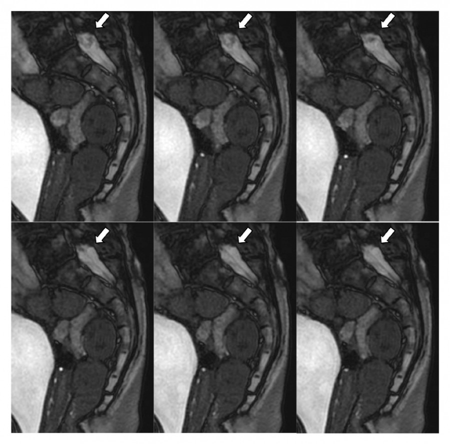
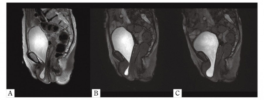
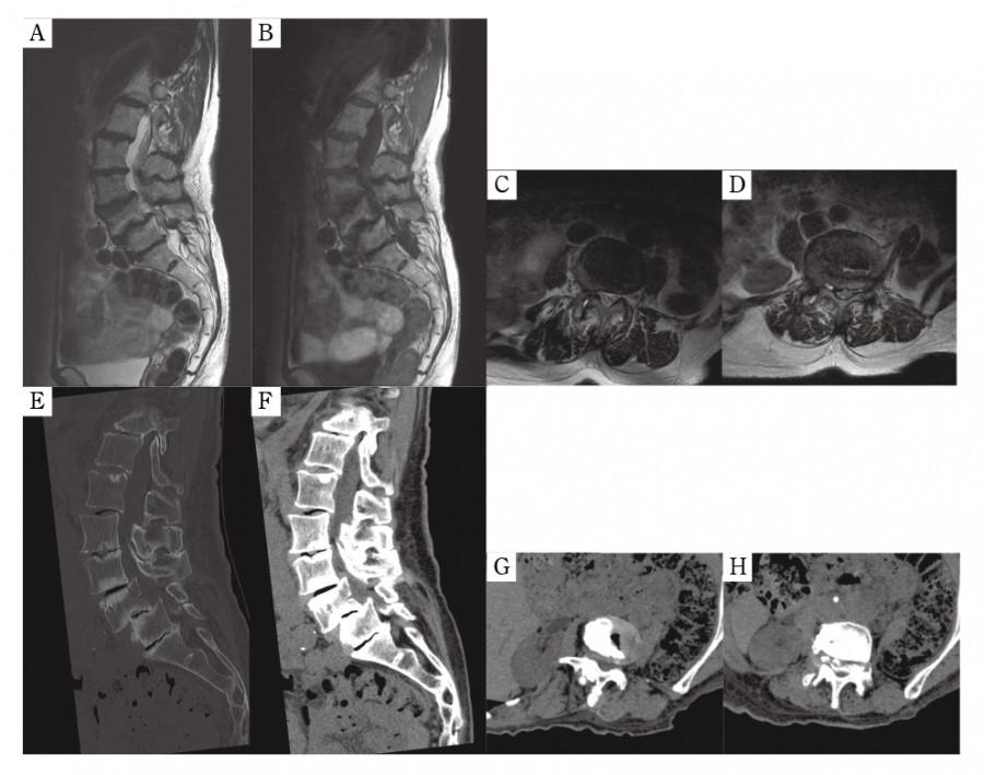
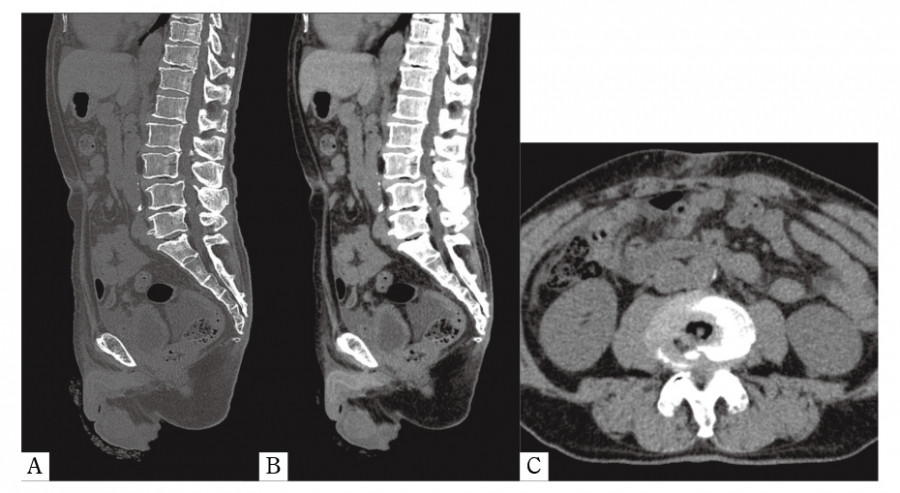
Case 1: A 74-year-old female with a history of LSS presented to our Department of Urology with frequent urination. The patient was diagnosed as having uterine and bladder prolapse. Pelvic cine MRI scan was conducted for detailed evaluation. While the Valsalva maneuver was performed to diagnose pelvic organ prolapses, we observed movement of the cauda equina. Spine MRI and CT, performed one year before presentation, showed severe LSS due to degenerative spondylolisthesis.
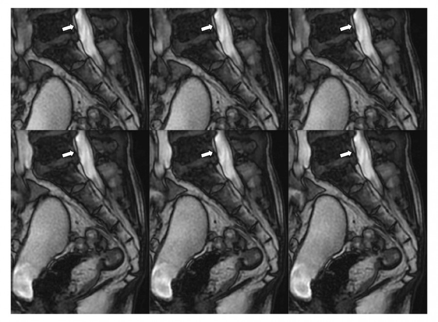
Case 2: A 73-year-old male underwent radical prostatectomy for prostate cancer. A follow-up cine MRI to confirm urethrorrhea showed the cauda equina moving during the Valsalva maneuver. Moderate LSS due to degenerative spondylolisthesis was retrospectively found on abdominal CT performed before prostatectomy.
Conclusion: The findings of our report suggest that movement of the cauda equina during the Valsalva maneuver may be implicated in LSS.
References
- 3.de Schepper EIT, Overdevest GM, Suri P, Peul WC, Oei EHG, Koes BW, et al. Diagnosis of lumbar spinal stenosis:an updated systematic review of the accuracy of diagnostic tests. Spine, 38:E469-481, 2013.
- 6.Koslosky E, Gendelberg D. Classification in Brief:The Meyerding Classification System of Spondylolisthesis. Clin Orthop Relat Res, 478:1125-1130, 2020.
- 7.Alsaleh K, Ho D, Rosas-Arellano MP, Stewart TC, Gurr KR, Bailey CS. Radiographic assessment of degenerative lumbar spinal stenosis:is MRI superior to CT? Eur Spine J, 26:362-367, 2017.
- 11.Drake RL, Vogl AW, Mitchell AWM. Gray’s anatomy for students 4th edition. Gray’s Anat students 4th Ed. Elsevier Inc, p104, 2019.
- 12.Groen RJM, du Toit DF, Phillips FM, Hoogland PVJM, Kuizenga K, Coppes MH, et al. Anatomical and Pathological Considerations in Percutaneous Vertebroplasty and Kyphoplasty:A Reappraisal of the Vertebral Venous System. Spine, 29:1465-1471, 2004.
- 15.Nathani KR, Naeem K, Rai HH, Barakzai MD, Iftikhar H, Khan SA, et al. Role of redundant nerve roots in clinical manifestations of lumbar spine stenosis. Surg Neurol Int, 12:218, 2021.
- 17.Srivastava A, Sood A, Joy SP, Woodcock J. Principles of physics in surgery:the laws of flow dynamics physics for surgeons — Part 1. Indian J Surg, 71:182-187, 2009.
Figures
Supplementary material
Supplemental Movie 1. Download (MOV)
A 74-year-old female with a history of lumbar spinal canal stenosis (Case 1). Cine MRI shows a large movement of the cauda equina in an inchworm-like manner during the Valsalva maneuver. The same image is shown in Figure 1.
Supplemental Movie 2. Download (MOV)
A 73-year-old male (Case 2). The cine MRI shows a small movement of the cauda equina during the Valsalva maneuver. The same image is shown in Figure 4.