The content of research paper

Recent topics in diagnosis and treatment of malignant spinal tumors

Yoshihiro Matsumoto

Author information
  • Yoshihiro Matsumoto

    Department of Orthopaedic Surgery, Fukushima Medical University

1. Topics in diagnostic imaging

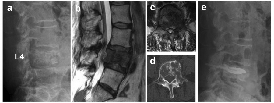
Spinal cord tumors are broadly classified into intramedullary, intradural, extramedullary, epidural, or spinal dumbbell tumors (SDT). SDT extend into or out of the spinal canal and have an hourglass-like appearance.


SDT, including schwannomas, are frequently benign tumors. However, PMST, such as malignant peripheral nerve sheath tumors (MPNST), can occur as malignant SDT (MSDT). Although challenging, MSDT can be cured with appropriate treatment at the localized stage. However, most MSDT proliferate rapidly and often cause neurological damage as they spread in the spinal canal.


Therefore, a prompt and accurate diagnosis is necessary. In fact, in many cases, histological analysis is required for a correct diagnosis, as there are many histologic types of MSDT. A biopsy is necessary for a definitive diagnosis; however, if the tumor is located in close proximity to adjacent vital organs such as blood vessels or the intestinal tract, it may require highly invasive maneuvers.


In contrast, benign SDT (BSDT) such as schwannomas have an indolent course, and resection can be avoided in such cases, which often discourages invasive biopsy. However, no method has been established to differentiate between BSDT and MSDT based on imaging. Therefore, we compared the imaging features of MSDT with those of BSDT and developed a simple scoring method to determine the benign or malignant status before surgery.


We included 59 patients with histologically confirmed SDT. The histological diagnoses and number of cases are shown in Table 1. A dumbbell scoring system (DSS) was developed and statistically analyzed by extracting the features of computed tomography (CT) and MRI images. The factors included in the DSS were: tumor diameter >5 cm (two points), indistinct borders (2 points), lobular morphology (1 point), and bone destruction (1 point), which were significantly more common in malignant tumors (Table 2). In each case, the scores were summed and the DSS score was calculated; the median DSS score was 0 for benign tumors and 5.5 for malignant tumors. Using 3 points as the threshold, differentiation between benign and malignant tumors was achieved with a sensitivity of 90% and a specificity of 84.6%. This indicated a high probability of differentiation between BSDT and MSDT1).


If malignancy is suspected by scoring, a definitive diagnosis can be made using CT-guided biopsy, which enables appropriate treatment planning, including consideration of additional preoperative chemotherapy and selection of a reliable surgical method to secure the resection margin. If the score is below the cutoff value, careful follow-up is also an option. However, the present analysis was a retrospective study with a limited number of cases, and future multicenter prospective validation is necessary to generalize the scoring system.


A new modality recently introduced is PET-MRI, which has begun clinical applications2). PET-MRI has several advantages over PET-CT. In pediatric imaging, multiple CT scans performed over a long period of time are known to increase the risk of developing secondary cancers3). PET-MRI, on the other hand, requires less radiation exposure than PET-CT, reportedly reducing the cumulative dose by 50-70% during follow-up for pediatric lymphatic tumors4).


In addition, MRI provides greater contrast, owing to differences in soft tissue organization, allowing for an accurate assessment of the extent of tumor extension. With this advantage, a report on limb-onset osteosarcoma found that the maximum standardized uptake values inside the tumor after preoperative chemotherapy were significantly correlated with the efficacy of chemotherapy5). A similar analysis was performed using PET/CT, but the results were not comparable with those obtained using PET-MRI6).


Furthermore, PET-MRI can identify tumor extension in the spinal canal and surrounding soft tissues of the paraspinal region, especially in the vicinity of neural tissue, and is useful in determining the extent of resection, radiation, and other treatment strategies. Thus, it may be effective for the diagnosis and treatment of PMST. However, studies on the usefulness of PET-MRI in PMST are scarce, and further validation is required.
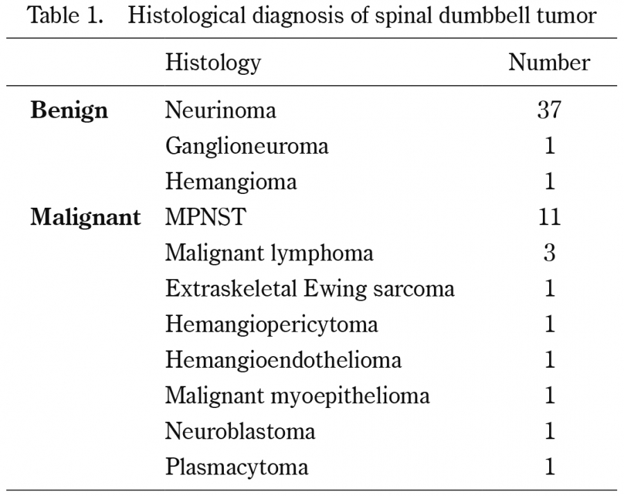


Table 1.

Histological diagnosis of spinal dumbbell tumor

MPNST: Malignant peripheral nerve sheath tumor
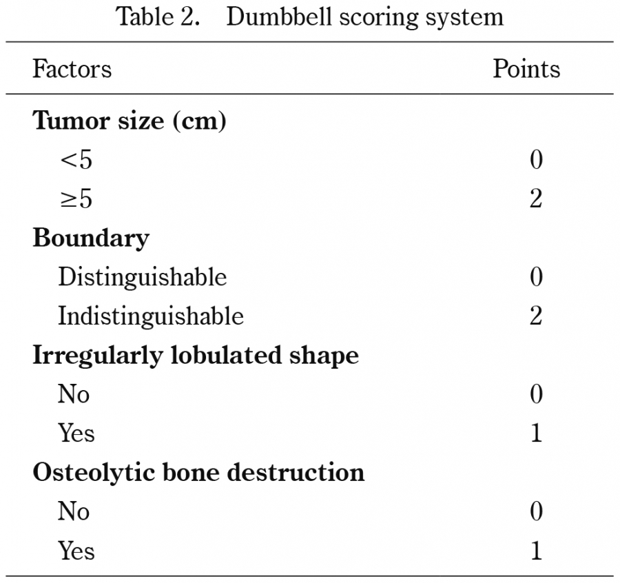


Table 2.

Dumbbell scoring system
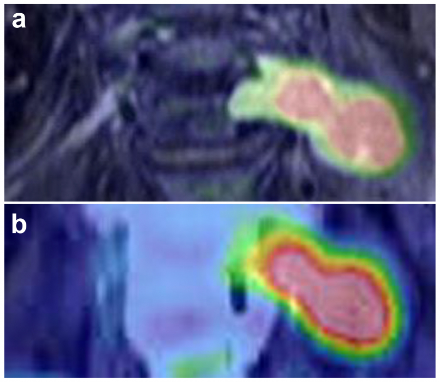
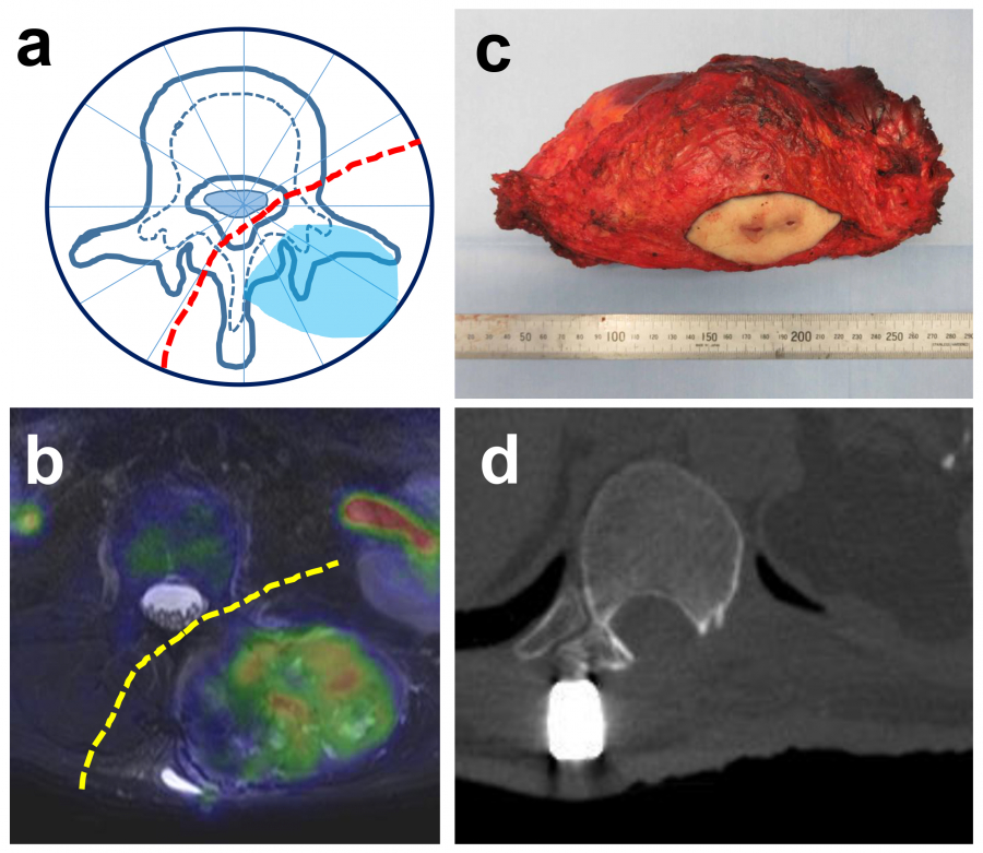
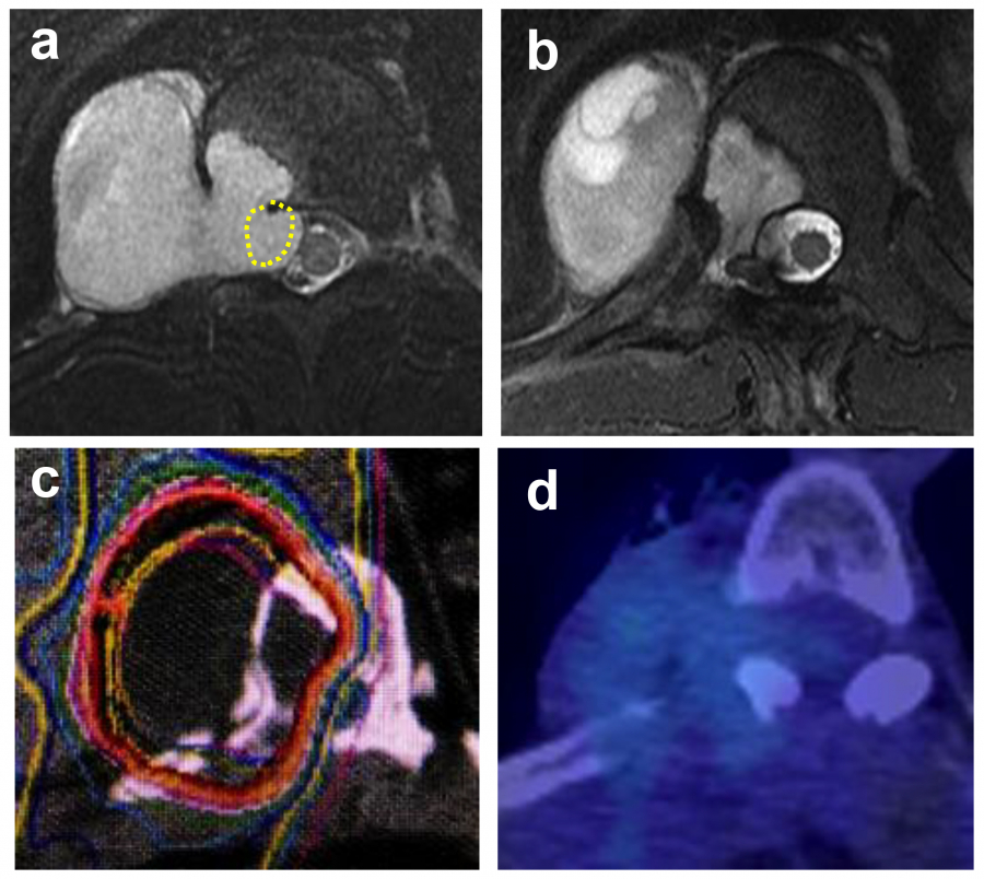


Fig. 1.

Positron emission tomography and magnetic resonance imaging (PET-MRI). (a) PET-MRI fusion image, (b) PET-computed tomography (CT) fusion image. Compared to PET-CT fusion images, PET-MRI fusion images demonstrate superior spatial resolution, clearly depicting tumors and surrounding tissues.

2. Improvements in radical resection for PMST

Surgical resection is the basic treatment for PMST, especially for tumors such as chondrosarcoma, which do not respond well to chemotherapy and radiotherapy. En bloc resection is performed using the Weinstein-Boriani-Biagnini surgical staging system (Fig. 2)7,8), depending on the localization defined by MRI or CT images. It is typically classified into three standardized methods:


Fig. 2.

Weinstein-Boriani-Biagnini (WBB) surgical staging system. The spine and spinal cord are divided into 12 counterclockwise regions and five layers (A to E, from prevertebral to dural involvement) to uniformly indicate tumor localization.


1) Vertebrectomy


This procedure is used when the tumor is localized in zones 4-8 or 5-9, i.e., when there is no tumor invasion in at least one of the pedicles. It is similar to a total en bloc spondylectomy (TES). Depending on the degree of anterior extension of the tumor, either a posterior-only or anterior-posterior approach is used. Vertebrectomy differs from TES in that the vertebral arch is resected piecemeal, particularly if the tumor has invaded the pedicle on one side. Fig. 3 shows a case in which vertebrectomy was performed according to TES.


Fig. 3.

Representative case of vertebrectomy. (a) A sagittal T2-weighted fat-suppressed MRI image of a 40-year-old male with T8 chondrosarcoma (Grade I). (b) Axial T2-weighted MRI image. (c) Macroscopic view of resected vertebra. (d) Postoperative X-ray of the thoracic spine (AP view).


2) Sagittal resection


This method is applied to eccentrically localized tumors (zones 2-5 or 8-11). For safe resection, a combined anterior and posterior approach is necessary, including the possibility of a joint resection involving multiple vertebrae and ribs. Specifically, the anterior approach is used first (extrapleural-retroperitoneal and retroperitoneal approaches are often used in the thoracic, thoracolumbar, and lumbar spines) to ligate the branching vessels from the major vessels, such as the intercostal and lumbar arteries, to the vertebral body, expand the vertebral body anteriorly and laterally, create a transverse groove in the vertebral body, and resect the intervertebral disc anteriorly. The ribs are amputated if necessary. The vertebral body, vertebral arch root, and transverse process are then broken off using a chisel while avoiding the dural canal, and the tumor is resected as a single mass. Fig. 4 shows a case of chondrosarcoma of the thoracic spine treated with sagittal resection.


Fig. 4.

Representative case of sagittal resection. (a) A 24-year-old female with T9 chondrosarcoma (Grade II), WBB2-4, showing tumors present in layers A, B, and C. Dashed line indicates the planned resection line. (b) T2-weighted MRI image. Dashed lines indicate the planned resection lines and arrows indicate the tumor. (c) Intraoperative gross findings, showing combined resection of the chest wall and vertebrae, with exposure of the dura mater and lungs. (d) Macroscopic view of resected tumor. (e) Postoperative axial CT image.


3) Posterior resection


Posterior resection is indicated when the tumor is located posteriorly, that is, counterclockwise from zones 3 to 10. If the tumor is located across the midline, the dural canal is developed above and below, the intervertebral joints are resected, and the bilateral vertebral arch roots are osteotomized and lifted for resection. If the tumor is slightly eccentric, the recently reported contralateral osteotomy of the pedicle and posterolateral elements for en bloc resection (COPPER) can be beneficial9). The COPPER approach involves resection of the healthy vertebral arch alone, and osteotomy is performed by inserting a chisel diagonally across the dural canal.


All the aforementioned resection methods are highly difficult operations, and to perform them safely and reliably, a detailed preoperative plan based on the biological characteristics of each tumor is necessary. To implement this plan, intraoperative navigation, ultrasonic bone scalpels, power devices for hemostasis and soft tissue handling, and other state-of-the-art surgical instruments are useful. Most importantly, a multidisciplinary team must be established in collaboration with various departments, including vascular, thoracic, abdominal, and plastic surgery.

3. Advent of heavy particle therapy

As noted earlier, the most important aspect of improving PMST outcomes is performing radical surgery at the initial visit. However, in practice, we often encounter cases where radical resection is impossible at the time of initial diagnosis. In such cases, radiotherapy is indicated.


However, PMST as well as bone and soft tissue tumors in the extremities are often radioresistant, and conventional radiation therapy is considered insufficiently effective. Therefore, a new type of radiation therapy, carbon-ion radiotherapy (CIRT), has been developed and applied to bone and soft tissue tumors10).


Heavy particles have an atomic number of 2 or higher, and carbon-ion beams are mainly used in CIRT. An important characteristic of heavy-particle beams is that they exhibit Bragg peaks. As a high-speed charged particle passes through matter, it gradually loses energy while causing ionization; however, just before it stops, it releases its maximum energy and then loses it. This phenomenon is called the Bragg peak, after its discoverer. The Bragg peak allows CIRT to deliver large doses of radiation to cancerous lesions, with little radiation beyond the target. Another feature of heavy-ion radiation is its high relative biological effectiveness, which is correlated with the amount of energy delivered per unit length, i.e., linear energy transfer (LET). It is expected to be effective in treating carcinomas resistant to radiotherapy11).


In an analysis of 47 PMST cases reported by the National Institute of Radiological Sciences, more than 90% of the patients received a total dose of 64 GyE or more in 16 fractions12). The median survival time was 44 months, with a 5-year local control rate of 79% and 5-year cumulative survival rate of 52%. One issue is the recurrence of marginal radiation fields near the spinal cord in patients with dural canal compression due to intraspinal tumor extension13).


The radiation dose tolerance of the spinal cord should be considered when treating tumors near it. Several reports have suggested that the tolerable dose for the spinal cord is approximately 50 GyE. If this dose is exceeded, it could lead to late radiation myelitis13), which in turn causes irreversible tetraplegia or paraplegia. Therefore, the irradiation dose must be maintained below 50 GyE in the vicinity of the spinal cord. On the other hand, 60-70 GyE is considered necessary for the local control of sarcomas, and it is highly likely that an insufficient dose on the spinal cord side leads to limbal recurrence. For this reason, CIRT was often avoided in cases of tumors with spinal cord compression, and many were shifted to palliative irradiation with conventional radiation.


To overcome this problem, we developed a new treatment system in which CIRT is performed after separation surgery (SS), in which only the area where the tumor contacts the spinal cord is removed (CIRT-SS) (Fig. 6). The SS provides a margin of several millimeters around the spinal cord and is expected to allow local control of the tumor while avoiding radiomyelitis. In a mid-term analysis, the 2-year local control rate in the initial treatment group was 87.5%, and no radiation myelitis was observed14), suggesting that CIRT-SS may be an effective treatment for PMST with spinal cord compression. However, the possibility of accelerated distant metastasis due to tumor curettage cannot be ruled out and further studies are needed to investigate this possibility.


A common adverse event associated with radiotherapy for bone tumors is pathological fracture after irradiation. Irradiation causes bone fragility and increases the risk of fractures. In particular, the frequency of post-irradiation vertebral fractures has been reported to be approximately 13.9%, with irradiation methods that concentrate high doses on the lesion, such as stereotactic radiotherapy. This is higher than the fracture incidence after conventional irradiation (3-5%)15).


Post-irradiation vertebral fractures were presumed to occur at a higher rate with CIRT than with other treatment modalities because of the high doses delivered to the vertebrae, similar to that with stereotactic radiotherapy. However, no such findings have been reported as yet. A review of 30 PMSTs treated with CIRT revealed that the frequency of post-irradiation vertebral fractures was as high as 23%, with a median time of occurrence of approximately 7 months16).


In CIRT, treatment planning is based on a simple CT. As metal artifacts prevent accurate treatment planning, implant fixation should be avoided prior to irradiation. Therefore, in cases where there is concern about vertebral fracture after irradiation, the patient should be carefully monitored in collaboration with a radiotherapist, and additional fixation should be considered at the appropriate time.

4. Development of novel drug therapies

Similar to malignant bone and soft tissue tumors of the extremities, chemotherapy-sensitive tumors such as osteosarcoma, Ewing sarcoma, and multiple myeloma are treated with drug therapy in combination with surgery or radiation therapy. Another major advancement in recent years has been the introduction of drug therapy for giant cell tumor of bone (GCTB).


GCTBs are characterized by marked bone destruction and account for approximately 5% of all primary bone tumors. Histologically, GCTB is classified as intermediate-grade and is composed of spindle-shaped quads and osteoclast-like giant cells, which are the main components of GCTB17). The epiphysis of the long tubular bone is the predominant site of occurrence; however, a few cases have also been reported in the trunk, including the spine and pelvis. Because of the local invasiveness of the lesion, some form of adjuvant therapy, such as phenol/ethanol treatment, is often combined with thorough curettage to prevent recurrence in cases of long bones. However, in spinal GCTB, it is difficult to add adjuvant therapy, and multiple relapses after curettage often occur, making treatment difficult.


Recently, receptor activation of nuclear factor κB ligand (RANKL) has been shown to be essential for osteoclast differentiation18). Since GCTB also contains many osteoclast-like giant cells, the effect of denosumab, a monoclonal antibody against RANKL, was investigated and confirmed for GCTB19). Denosumab efficacy has also been recognized in Japan20) and covered by insurance since 2014. As shown in Fig. 7, it represents a major breakthrough in the treatment of spinal GCTB, which has been difficult to treat until now.


However, the widespread use of denosumab for GCTB treatment has made several concerns apparent, the most significant being the optimal treatment duration. Denosumab-treated tissue specimens show more than 90% osteoclast-like giant cell loss and irregular osteosclerosis20). However, spindle-shaped cells remain in the stroma and osteosclerotic nests, making it difficult to determine whether these cells are GCTB tumor cells or normal osteoblasts. It is difficult to determine whether these cells were GCTB tumor cells or normal osteoblasts.


This question was answered by elucidating a tumor cell-specific genetic mutation in GCTB. The H3F3A mutation (H3.3G34W), encoding a histone 3.3 (H3.3) mutant protein, is a driver gene mutation specific to giant cell tumors. H3.3G34W mutant protein is found in approximately 90% of giant cell tumors21) and can be easily identified by immunostaining, serving as a marker for GCTB tumor cells. In our study, numerous H3.3G34W stain-positive spindle-shaped cells remained in the stroma and osteosclerotic nests of the tissue specimens after denosumab administration22) (Fig. 9).


Based on these results, GCTB tumor cells were expected to persist even after denosumab administration, and upon discontinuation of the medication, tumor regrowth was considered inevitable. Therefore, surgical resection is necessary in order to discontinue treatment. However, excessive bone sclerosis has been reported following long-term treatment with denosumab, making surgery difficult for vertebral GCTB23). Further case series are needed to clarify the appropriate administration of denosumab for difficult-to-treat GCTB and its combination with surgery.


Fig. 5.

Representative case of posterior resection. (a) A 57-year-old male with dedifferentiated liposarcoma arising from the paraspinal region of the lumbar spine, with tumors present in WBB1-3, layers A, B, and C, (b) Axial T2-weighted fat-suppressed MRI image. Dashed lines indicate the planned resection line. (c) Macroscopic view of resected tumor. (d) Postoperative axial CT image.


Fig. 6.

Carbon-ion radiotherapy surgical support (CIRT-SS) for primary malignant spinal cord tumors. a) A 49-year-old male with T11/12 malignant peripheral nerve sheath tumor causing dural compression (dashed lines). b) Decompression of the dura mater by separation surgery (excision of dashed lines). c) CIRT-SS enabling sufficient irradiation dose to the entire tumor (red line indicates 90% dose). d) Post-operative PET-CT axial image. FDG uptake of the tumor is not shown.


Fig. 7.

Example of denosumab administration for giant cell tumor of bone arising as a spine tumor. (a) A 40-year-old female with cervical vertebral giant cell tumor of bone. (b) Histological image of biopsy specimen showing numerous giant cells (arrows); hematoxylin and eosin (HE) stain, × 100. (c) Axial T2-weighted fat-suppressed MRI image. (d) PET-CT axial image. (e, f) Axial CT image approximately 6 months after denosumab administration. (e) PET-CT showing tumor shrinkage and bone formation within the tumor. (f) Confirming reduced tumor activity.


Fig. 8.

Histological images of giant cell tumor of bone before and after denosumab administration. (a, b) Images before administration:(a) HE stain. (b) Immunostaining using anti-H3.3G34W antibody. (c, d) Images after administration:(c) HE stain showing significant bone formation and detachment of giant cells. (d) Immunostaining using anti-H3.3G34W antibody revealing numerous anti-H3.3G34W antibody-positive cells surrounding and within the formed bone. (a-d) × 200.


Fig. 9.

Posterior fixation surgery using percutaneous pedicle screws (PPS) for metastatic spinal tumor (MST). An 82-year-old male with multiple spinal metastases from leiomyosarcoma. (a) Sagittal T2-weighted fat-suppressed MRI image. (b, c) Multilevel posterior fixation surgery using PPS:(b) Postoperative AP X-ray. (c) Postoperative lateral X-ray.

5. Advances in MST treatment

(1) Widespread use of Bone Metastasis Cancer Board (BMCB)


The prognosis of malignant tumors has steadily improved owing to advances in treatment methods, such as the widespread use of molecular-targeted drugs and the emergence of tumor immunotherapy. Therefore, although the prognosis of bone/spine metastases was previously considered to be within a few months, patients often survive for up to a year depending on the histological type.


Bone metastases, especially MST when symptomatic, cause intractable pain and paraplegia, reducing the quality of life (QOL) and overall performance status (PS). As a result, continuing treatment for the primary tumor may become difficult and prognosis may worsen. However, unsatisfactory treatment for MST is not uncommon, mostly owing to a lack of awareness in primary departments and the often non-aggressive approach of general orthopedic surgeons.


To improve this situation, organization of so-called BMCBs, where multiple disciplines collaborate at multiple facilities to diagnose and treat metastatic bone tumors as a single hospital, has been increasingly adopted in recent years. In a BMCB, each primary department, including orthopedic surgeons (spine surgeons), radiation therapists, rehabilitation physicians, medical oncologists, palliative care members, physiotherapists, nurses, and social workers discuss treatment policies for patients with bone metastasis from various perspectives and formulate multidisciplinary treatment plans. BMCBs are expected to increase patient referrals to hospitals and the number of MST surgeries, and are recommended by guidelines for bone metastasis treatment24).


(2) Introduction to minimally invasive spine stabilization (MISt) for MST


Indications for surgical intervention for MST include:1) progressive paralysis, 2) impending paralysis due to spinal instability, and 3) pain that is poorly controlled by drugs and radiotherapy. On the other hand, surgery is generally considered to be indicated only for patients with a prognosis of at least 6 months. This is because surgery aimed at improving QOL may in fact worsen the patient’s general condition and prognosis, as it is an invasive procedure for patients with MST whose biological reserve is originally compromised.


However, with the recent development of MISt, the indications for MST surgery have changed. Percutaneous pedicle screws (PPS) and percutaneous kyphoplasty (BKP) are the most useful MISt techniques for MST and are outlined as follows:


(a) Application of PPS to MST


In PPS, a guidewire is inserted under fluoroscopic guidance through a small skin incision, and a hollow vertebral root screw is inserted along the wire. The advantages of PPS include:(1) minimal invasiveness with minimal blood loss ;(2) rapid wound healing, allowing postoperative radiotherapy and chemotherapy to begin early ;(3) reduced seeding of tumor cells by the surgical procedure ;and (4) short procedure times. Its disadvantages include:(1) difficulty in bone grafting and a possibility of implant failure when long-term prognosis is achieved;(2) technical difficulty in obtaining adequate fluoroscopic images proximal to the upper thoracic spine ;(3) insufficient decompression through small incisions.


However, the advantages often greatly outweigh the disadvantages, especially in cases with multiple spinal lesions. The technique can be applied in cases where surgery using conventional methods must be avoided (Fig. 9).


(b) Experience with BKP in MST


Destruction of the vertebral body associated with tumor invasion causes instability and significant pain. In such cases, pain relief by radiotherapy is limited, immobilization is difficult in the absence of paralysis, and pain control is poor even with the use of high-dose narcotic analgesics in many cases. However, tumor-related vertebral fractures often show more severe vertebral destruction than osteoporotic vertebral fractures, and the risk of leakage into or out of the spinal canal during cement injection is high.


BKP, a cemented vertebral body augmentation technique, has been indicated for vertebral body fractures associated with multiple myeloma or MST in up to three vertebral bodies in Japan since December 2011. BKP was considered safer than vertebroplasty because a balloon is first expanded within the vertebral body to provide space for cement filling before injection27).


The Cancer Patient Fracture Evaluation (CAFE) study reported the highest level of evidence for BKP in tumor-related vertebral fractures. In this study, 117 patients with MST were randomly assigned to the BKP (65 patients) and control (conservative treatment; 52 patients) groups. The results showed that significant pain improvement was achieved only in the BKP group, and this effect persisted for 12 months after the start of treatment. Furthermore, no serious adverse events associated with cement leakage were observed, indicating that BKP is an effective and safe treatment option for MST28).


However, reports on the use of BKP for MST in Japan have been few. We have been actively performing BKP for tumor-related vertebral fractures without paralysis (Fig. 10). In our experience with 17 consecutive patients, pain on the Numerical Rating Scale improved from 7±2.3 (SD) preoperatively to 2.2±2.1 at 1 month postoperatively, and 1.8±1.9 at the final observation. This indicates successful replication of the CAFE study results. However, one case presented with a crushed vertebra following BKP, forcing the addition of posterior fixation with PPS. Further study is needed to determine the best indication for this procedure.


(3) Paradigm shifts in the treatment system for MST


Surgical intervention has been reported to improve PS and QOL in patients with MST and to prolong life expectancy, as chemotherapy is often administered after surgery29). Therefore, the appropriate utilization of BMCB and MISt may expand the indications for surgery in patients with frailty and improve outcomes. While surgery has been a “wait-and-see” procedure, whereby patients are considered for surgery only after a request is received from the respective primary department, in the future, prophylactic surgery using MISt will be considered for cases of progressive spinal instability or tumor growth.


The treatment system for MST is currently being updated. The focus is shifting towards preventing the deterioration of QOL and PS due to MST, continuing outpatient chemotherapy, and enabling patients to remain at home, which may become mainstream in the future.


Fig. 10.

Balloon kyphoplasty (BKP) for MST. (a) Lateral X-ray image of a 57-year-old female with L4 metastasis from breast cancer. (b) Sagittal T2-weighted MRI image. (c) Axial T2-weighted MRI image. (d) Axial CT image showing intact posterior wall of the spinal canal. (e) Post-BKP lateral X-ray image.

Conclusion

The knowledge and skills required for the diagnosis and treatment of malignant spinal tumors are extremely diverse, and multidisciplinary treatment must be practiced in collaboration with other departments. As described in this paper, there have been remarkable advances in the field of both primary and metastatic malignant spinal tumors, and further improvements in treatment outcomes are expected in the future.

Acknowledgements

This work was partially supported by a Grant-in-Aid for Scientific Research from the Promotion of Science (# 24K02569) and the Japanese Orthopaedic Association (JOA, Subsidized Science Project Research (2022-1). Editage provided English language editing.

Conflict of Interest Disclosure

The author declares no conflict of interest.