The Department of Nuclear Medicine Physics conducts diagnostic imaging and clinical research on various diseases using nuclear medicine imaging devices such as PET/CT, PET-MRI, and SPECT/CT systems. Nuclear medicine imaging is an examination in which a radiopharmaceuticals are administered into the body and various biological functions are visualized by detecting radiation. Nuclear medicine imaging analysis enables us to improve diagnostic techniques and evaluate absorbed doses from radiation, contributing to the promotion of research on highly accurate diagnostic and therapeutic methods.
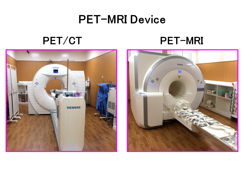
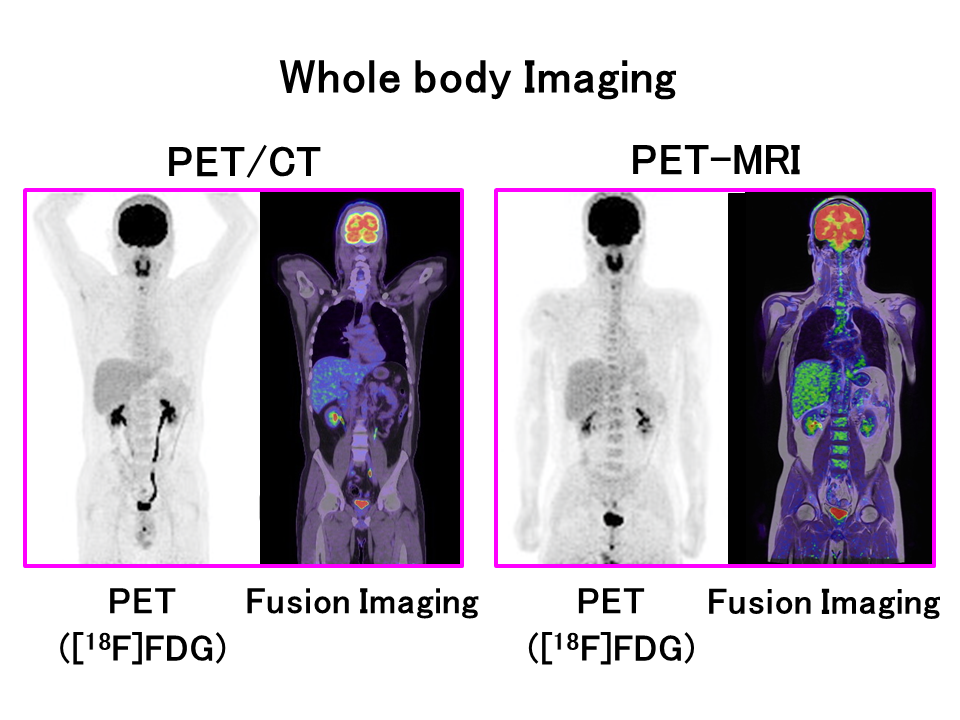
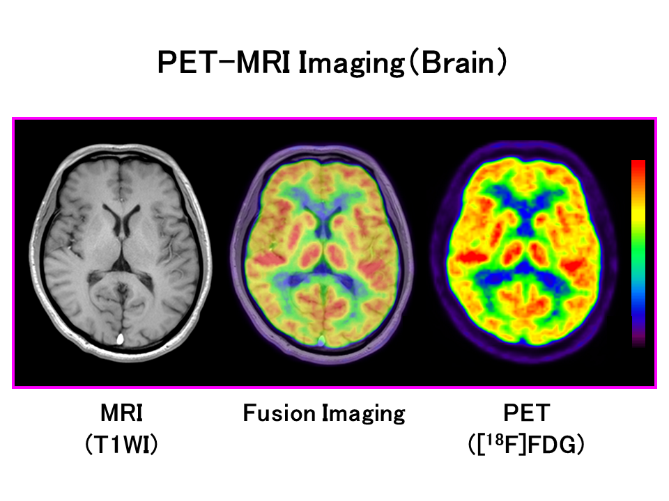
Nuclear medicine physics research will be conducted in collaboration with the University Hospital. One PET/CT scanner and one PET-MRI scanner have been in operation at the University Hospital since 2013, and one SPECT/CT scanner was installed in FY2025.The PET-MRI scanner is a hybrid type of diagnostic imaging system that incorporates a PET scanner into an MRI scanner, a diagnostic imaging system. PET-MRI can easily and accurately obtain fusion images of high-precision morphological images by MRI and biofunctional images by PET, and is very useful for diagnostic imaging and clinical research. In addition, SPECT/CT is used to image therapeutic radionuclides such as 177Lu and 211At to evaluate radiation dosimetry, which enables us to estimate treatment effects and side effects.
![先端臨床研究センター[ふくしま国際医療科学センター]](https://www.fmu.ac.jp/home/acrc/en/wp/wp-content/themes/sentan2%20_en/images/site-id.png)