The cintent of reseach paper

Investigation of biomarkers in a rare case of fulminant necrotizing enterocolitis in a preterm infant

Toshihiko Nakamura, Shota Inoue, Kana Ito, Eisuke Fukama, Tomoaki Nomura, Daisuke Hatanaka, Michiko Kusakari, Hidehiro Takahashi, Shingo Yamada

Author information
  • Toshihiko Nakamura

    Department of Neonatology, Japanese Red Cross Musashino Hospital

  • Shota Inoue

    Department of Neonatology, Japanese Red Cross Musashino Hospital
    Department of Pediatrics, National Defense Medical College

  • Kana Ito

    Department of Neonatology, Japanese Red Cross Musashino Hospital
    Department of Pediatrics, National Defense Medical College

  • Eisuke Fukama

    Department of Neonatology, Japanese Red Cross Musashino Hospital

  • Tomoaki Nomura

    Department of Neonatology, Japanese Red Cross Musashino Hospital

  • Daisuke Hatanaka

    Department of Neonatology, Japanese Red Cross Musashino Hospital

  • Michiko Kusakari

    Department of Neonatology, Japanese Red Cross Musashino Hospital

  • Hidehiro Takahashi

    Department of Neonatology, Japanese Red Cross Musashino Hospital

  • Shingo Yamada

    Shino-Test Corporation

Introduction

The cause of common necrotizing enterocolitis (NEC) is, against the background of an immature intestinal tract, considered to be related to such factors as the dose of enteral nutrition, its timing of administration, and the type of nutrition (whether breastfeeding or formula). Highly immature intestinal mucosa is prone to develop mucosal damage and tissue damage due to impaired blood circulation1). In addition, complications such as bacterial infections can lead to severe damage of the intestinal tract or necrosis2).


We encountered a case of infant death that could not be explained solely by the course of sepsis triggered by common NEC. A few cases of NEC are fulminant and death is remarkably rapid with diffuse intestinal necrosis seen on laparotomy or autopsy. Fulminant necrotizing enterocolitis (F-NEC) is defined as a death ascribed to NEC, with a period between diagnosis of NEC and death of less than 48 hours. However, the pathophysiology, characteristics, and prognosis of F-NEC are not clear. Endogenous damage-associated molecular patterns (DAMPs) are now known to be released from cells in response to infection, tissue injury, and/or necrosis. We report on the comprehensive evaluation of acute-phase reactants, cytokines, several cytotoxic markers such as high mobility group box-1 (HMGB-1), histone H3, and syndecan-1 as the basis for our diagnosis of F-NEC. In doing so, we retrospectively examine the extent of this disease, which resulted in death due to rapid deterioration of the infant’s general condition.

Case report

The baby, who had a gestational age of 30 weeks 2 days and birth weight of 1,097 g (3.1 percentile) was born by emergency cesarean section for the indication of non-reassuring fetal status. INSURE-therapy (Intubation, Surfactant, Extubation) was performed for respiratory distress syndrome and then nasal CPAP was started for his respiratory care. Enteral nutrition was very carefully started from day 2 of life, and the amount was gradually increased mainly with breast milk. Enteral administration of probiotics was started from day 1. Gastric residue remained almost non-existent in the pre-enteral nutrition check. At the age of 14 days, the infant’s condition suddenly deteriorated shortly after vomiting a large quantity of bile-free fluid. After a sepsis work up, administration of cefmetazole sodium and vancomycin hydrochloride was started intravenously. Intestinal peristaltic sounds had diminished 4 hours later and disappeared by 16 hours, and abdominal distension had become marked, at which time the skin color of the entire abdomen changed to dark red. Artificial respiration management was begun with the administration of γ-globulin and catecholamine, but a decrease in blood pressure was observed from around 12 hours and his heart rate exceeded 200/min. He was diagnosed to be in a state of shock with a progressively increasing metabolic acidosis with hyperlactatemia. The DIC score for infants was 5 points and DIC was confirmed3). Escherichia coli were isolated by blood culture at 24 hours, but the catheter tip culture was negative. The patient was diagnosed as having endotoxin shock due to the bacterium, and exchange transfusion was performed with the expectation that the endotoxin and inflammatory cytokines would be removed. However, his general condition showed no signs of recovery, and he died 42 hours after the diagnosis of NEC stage II (Fig. 1-A,-B, Table 1).

Fig. 1-A. Transition of clinical symptoms and treatment after onset:

The time of onset of vomiting was set to 0 and displayed over time. → in the abdominal radiograph refers to the gas in the portal vein. The bubble gas pattern observed early after onset spread from the lower right half to occupy the entire lower half of the abdomen. Throughout the course, no free air was found in the abdomen. The blood flow in the celiac artery (CA) and superior mesenteric artery (SMA) was followed by abdominal echo. The SMA could be visualized at onset, but visualization of the CA was not possible. We noted that the blood flow velocity of the celiac artery, which seems to be compensatory, was increased. Twenty hours after onset, the abdominal wall rapidly increased in fullness, and the color of the abdominal wall became dark purple instead of inflammatory red.


Fig. 1-B. Transition of clinical vital signs after onset:

The time axis is the same as in Fig. 1-A. From the vital signs 12 hours after the onset, it can be seen that the infant had gone into shock. After that, despite all the possible treatments, there was no recovery from shock.


Table 1. Laboratory data four hours after the onset of gastrointestinal signs.

Material and methods

With the informed consent of his parents, residual serum was used to create cytokine profiles and the measure cytotoxic markers HMGB-1, histone H3, and syndecan-1. Our investigation was approved by the Research Ethics Committee of our hospital. The parents of the infant were informed of the study design, and their written informed consent was obtained for submission of this case report.


Cytokine profiles of IL-1β, -6, -8, -10, TNFα, and G-CSF were assessed with an ELISA kit manufactured by R & D Systems. HMGB-1 measurements were performed by using a commercially available ELISA kit (Shino-Test Corp., Sagamihara, Japan). The detection sensitivity of this assay system was 0.2 ng/mL4). Because there is no commercially available ELISA kit, the measurement of histone H3 was performed by an ELISA prepared in our laboratory. The detection sensitivity of this assay system was 2.0 ng/mL5). Syndecan-1 was also measured by the ELISA method originally described by our laboratory6). Among the acute phase reactants (APR) CRP, α1-acid glycoprotein (AGP), and haptoglobin (Hp) were measured by LatessierTM (Shino-Test Corp.) using a turbidimetric immunoassay7).

Results

The transition of the APR was not significantly increased at 4 hours after the onset of gastrointestinal symptoms (Table 2), but CRP and AGP were remarkably increased at 12 hours after onset. Hp was below sensitivity from the beginning to end. CRP and AGP decreased once after the exchange transfusion but then increased to almost the previous value by 12 hours after the transfusion (Fig. 2).


IL-8 and IL-10 could not be measured at 5 hours after the onset because the sample volume was inadequate. However, the other four cytokines, (IL-1β, -6, TNFα, G-CSF), showed a significant increase at 5 hours after onset and then continued to increase. Although IL-1β and TNFα increased, the degree of increase of IL-6 and G-CSF was considerable compared to that of IL-1β and TNFα. IL-1β, TNFα, and IL-10 decreased once following the exchange transfusion, but all of them increased again to above their previous values in a short time. Rather, IL-6, IL-8 and G-CSF did not decrease.


With the exception of HMGB-1, there are no reference values for histone H3 and syndecan-1 in normal newborns4). However, due to the nature of these substances, it is hypothesized that the effect of perinatal hypoxic stress at 2 weeks after birth would be less than that of the acute period. Under this hypothesis, HMGB-1 and histone H3 were significantly increased 24 hours after onset5). Although syndecan-1 was also increased, it did not reach a level as high as its usual increase during adult sepsis, and it remained lower than the value seen in sepsis without disseminated intravascular coagulation6) (Fig. 3).

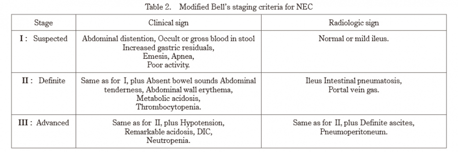
Table 2. Modified Bell’s staging criteria for NEC


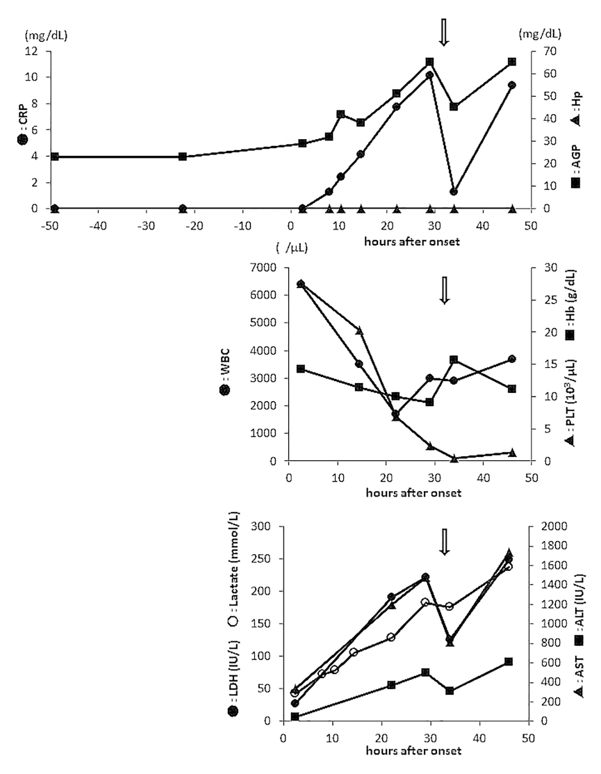
Fig. 2. Time course of acute phase reactants, peripheral blood cells, and releasing enzymes before and after onset:

Haptoglobin (Hp) remained negative from beginning to end. C-reactive protein (CRP) became positive 12 hours after the onset. The α1-acid glycoprotein (AGP) did not increase prior to CRP during feeding intolerance as has been previously reported. After onset, it increased slightly later than the increase in CRP. A decrease in these two acute phase proteins was seen once after exchange transfusion, but levels returned to the previous value in about half a day. White blood cell count and platelet count dropped sharply after onset. Hemoglobin did not decrease as much as the former two. Three of the releasing enzymes also increased after onset, did not increase to the same degree. Arrows mean blood exchange transfusion.


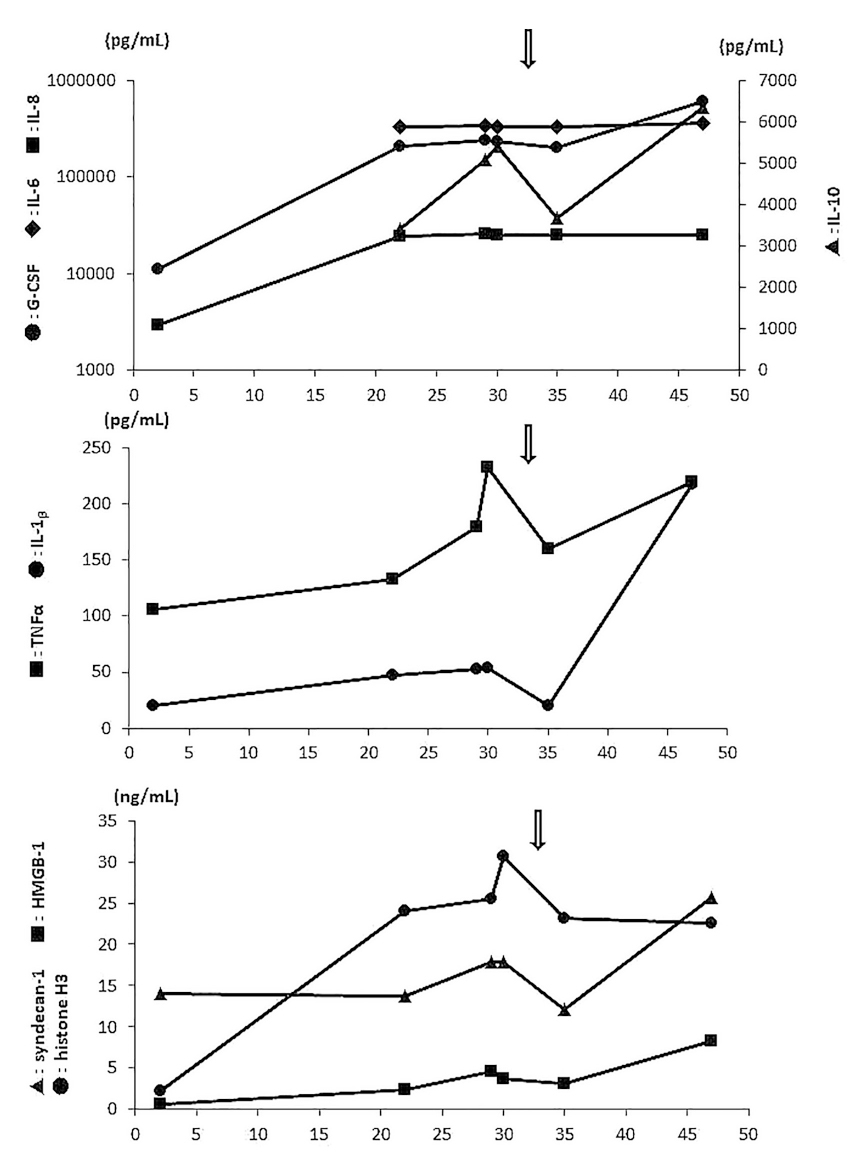
Fig. 3. Serial changes of cytokine profiles and the three cytotoxic markers after onset:

It can be seen that all six cytokines show high levels, and in particular, IL-6,-8,-10 and G-CSF have markedly elevated levels. Although these cytokines temporarily decreased after exchange transfusion, exacerbation exceeding the previous value was observed in about half a day. Considering the age of the newborn at onset, it is judged that the effect of ischemia-reperfusion injury at birth has disappeared. Based on this hypothesis, it is judged to be abnormal when these cytotoxic markers are present in the blood after onset. Considering adult reports, high mobility group box-1 (HMGB-1) and histone H3 are clearly elevated, but syndecan-1 is not significantly elevated. Serum syndecan-1 concentration in sepsis remains at a level that does not cause disseminated intravasclular coagulopathy. The arrows indicate blood exchange transfusion.

Discussion

The pathophysiology of NEC suggests that intra-luminal bacteria disrupt and invade the premature intestinal epithelium at the tips of intestinal villi. Endotoxin from these bacteria binds to Toll-like receptor 4 found on the intestinal epithelial cells, which facilitates the breakdown of the gut barrier and allows bacterial translocation9-12). As a result, it is quite possible that sepsis will occur as a complication. This process subsequently leads to an intense inflammatory response in the lamina propria mediated by TNFα, IL-1β, and other inflammatory cytokines. Then leukocytes and platelets adhere to the endothelium, preventing blood flow in the microvascular structure of the small intestine and lead to tissue deterioration1,2).


We judged this case to be consistent with F-NEC from the downhill course following onset. There are only occasional reports of F-NEC13-18). Its etiology, such as whether F-NEC is a subtype of NEC, has been little studied, but there are reports reviewing common points of the clinical symptoms. A reduction in the number of leukocytes, neutrophils and lymphocytes, a decrease in platelets, and an increase in the ratio of immature to mature cells in the leukocyte fraction are significant13-15). These findings are also consistent with those of the present case.


The cytokine profile of the infant was characterized by increases in TNFα and IL-1β that were about an order of magnitude lower than those seen during common neonatal sepsis19-21). However, IL-6, IL-8, G-CSF, and the anti-inflammatory cytokine IL-10 showed quite significant elevations. HMGB-1, histone H3, and syndecan-1 were elevated but not as high as reported in adult cases of sepsis7,8,22,23). Despite TNFα and IL-1β belonging to the inflammatory cytokines in the cytokine profile of the present infant, their elevations were minor, but marked increases in IL-6 and IL-8 were observed. Increases in IL-6 and IL-8 have been noted in the cytokine profiles of patients with NEC24-27) and neonatal asphyxia28-30). Common to both diseases is the presence of ischemic lesions at the site of injury.


Despite the notable increases in CRP and AGP, Hp remained below the measured sensitivity, which is also a characteristic trend of APRs. We have examined the involvement of cytokines in the production of APRs and found that Hp production required a sufficient increase in IL-1β. Although this patient’s IL-1β rose above the baseline level, it changed very little compared to the large rises in IL-6 and IL-8. The mechanism for this difference of elevation in IL-6 and IL-8 versus that of IL-1β is unknown, and elucidation of its mechanism may help to clarify the pathophysiology of F-NEC.


The examination of this case suggested that F-NEC is clearly different from common NEC in terms of the cytokine profile and the decrease in peripheral blood cell components. This difference cannot be explained simply by a difference in severity, and it seems urgent to carry out additional tests in similar cases. As far as the case is concerned, it is characterized by the presence of extraordinary hypercytokinemia and extremely poor clearance of these cytokines even after blood exchange transfusion. This patient may have a constitution in which the biological defense mechanism overreacts. To achieve this, because we fear that research performed at a single facility may take at least 10 years, it is urgent to conduct joint research among multiple facilities.

Conflict of interest disclosure

The authors have no conflicts of interest to declare.

Contributors

All authors were involved in the clinical care of the infant and contributed to the conception, drafting, review, and revision of the manuscript. All authors have read and approved the final version of the manuscript and take full responsibility for the work.