The cintent of reseach paper

A case of spontaneous parasitic myoma in a patient without a history of myomectomy treated laparoscopically

Hyo Kyozuka, Toki Jin, Misa Sugeno, Katsunori Kuratsune, Hiroki Ando, Fumihiro Ito, Hajime Odajima, Daisuke Suzuki, Yasuhisa Nomura

Author information
  • Hyo Kyozuka

    Department of Obstetrics and Gynecology, Ohta nisinouchi Hospital

  • Toki Jin

    Department of Obstetrics and Gynecology, Ohta nisinouchi Hospital

  • Misa Sugeno

    Department of Obstetrics and Gynecology, Ohta nisinouchi Hospital

  • Katsunori Kuratsune

    Department of Obstetrics and Gynecology, Ohta nisinouchi Hospital
    Department of Obstetrics and Gynecology, School of Medicine, University of Occupational and Environmental Health

  • Hiroki Ando

    Department of Obstetrics and Gynecology, Ohta nisinouchi Hospital

  • Fumihiro Ito

    Department of Obstetrics and Gynecology, Ohta nisinouchi Hospital

  • Hajime Odajima

    Department of Obstetrics and Pathology, Ohta nisinouchi Hospital

  • Daisuke Suzuki

    Department of Obstetrics and Gynecology, Ohta nisinouchi Hospital

  • Yasuhisa Nomura

    Department of Obstetrics and Gynecology, Ohta nisinouchi Hospital

Introduction

Uterine myomas, also known as leiomyomata, fibroids, fibromyomas, leiomyofibromas, and fibroleiomyomas, are benign tumors that originate from the clonal proliferation of smooth muscle cells in the uterus. They are common among women of reproductive age, with a prevalence of 20-60%1). Symptoms of uterine myomas include dysmenorrhea, hypermenorrhea, pelvic pressure, frequent urination, and sterility2). The prevalence of uterine myomas is approximately 6.6% in pregnant women in Japan3), representing a potential risk for preterm birth1). With advances in minimally invasive surgery, laparoscopic myomectomy has been extensively utilized in treating fibroids. During laparoscopic surgery, power morcellation devices are used to remove sizable specimens through small incisionsHowever, dissemination of benign tissues is an increasing concern, and sequelae have been reported4,5).


Spontaneous parasitic myoma (PM) is extremely rare because most reported cases are iatrogenic and linked to the unintentional seeding of fragments during previous laparoscopic myomectomy by morcellators6). Herein, we report a case of a spontaneous PM treated laparoscopically. The woman was of reproductive age and had no previous surgical history.

Case presentation

A 40-year-old Japanese multiparous woman presented to our hospital with heavy menstrual bleeding. She had no remarkable medical or previous surgery history. Although blood tests showed no evidence of anemia, magnetic resonance imaging (MRI) showed uterine myomas measuring 6.6 cm × 5.9 cm and 5.8 cm × 4.0 cm in size at the anterior and posterior walls, respectively (Figure 1). Therefore, a uterine myoma was diagnosed. Since the patient did not wish for further pregnancy, she underwent oral gonadotropin-releasing hormone antagonist therapy and was scheduled for total laparoscopic hysterectomy (TLH). Under general anesthesia, with the patient placed in the lithotomy position, trocars were positioned at the standard sites to avoid injury to the uterus Laparoscopy revealed an over fist-sized uterus due to the myoma occupying the pelvic cavity, and a TLH was performed. During the TLH, we found a thumb-sized tumor on the left side of the peritoneum (Figure 2) that was resected and collected from the left side port. The estimated total blood loss during the operation was 25 g. The patient was discharged on postpartum day 3 without complications. Histopathological examination of the resected tumor revealed a typical microscopic view of a benign leiomyoma, with sparse mitosis and no necrosis or marked atypia in the uterus. It also revealed that the resected tumor on the left side of the peritoneum showed evidence of leiomyoma without atypia (Figure 3a). Immunohistochemistry showed that the tumor cells were positive for smooth muscle actin (SMA), estrogen receptor (ER), and progesterone receptor, suggesting monoclonal tumor arising from the uterine smooth muscle tissue (Figure 3 b, c, d).


We retrospectively reviewed the MRI scans for lesions in this case and found the presence of a 1.8 × 1.0 cm low-density solid tumor in the left peritoneum, separate from the uterine and uterine myoma on T2-weighted MRI (Figure 4). Accordingly, we diagnosed the patient with a uterine myoma and a naturally occurring parasitic myoma.


Fig. 1. Sagittal T2-weighted MRI

Magnetic resonance imaging (MRI) showing enlarged uterine myomas that occupy the pelvic cavity (arrow).


Fig. 2. Intraoperative laparoscopic image

After laparoscopic hysterectomy is completed, we identified a thumb-sized tumor on the left side of the peritoneum (arrow). The patient had no previous surgical history.


Fig. 3. Pathological findings for the resected tumor of the left side peritoneum.

Hematoxylin and eosin, ×200 objective. Typical microscopic view of a benign leiomyoma, with no necrosis and without marked atypia (3a). The immunohistochemical evaluation of the resected tumor is positive for smooth muscle actin (3b), estrogen receptor (3c), and progesterone receptor (3d).


Fig. 4. Coronal T2-weighted MRI

After laparoscopic surgery, we reviewed the MRI scans taken before surgery. T2 weight MRI lesion indicates the presence of a 1.8 × 1.0 cm low-density solid tumor in the left peritoneum, apart from the uterine and uterine myoma (arrow).

Discussion

Uterine smooth muscle tumors with unusual growth patterns include three primary neoplasms:intravenous leiomyomatosis (IVL), benign metastasizing leiomyoma (BML), and leiomyomatosis peritonealis disseminata (LPD)7). IVL is defined as the pathological presence of smooth muscle cells growing in venous or lymphatic vessel spaces without invading them7). The plausible etiology of IVL comprises uterine leiomyomas entering the venous system or directly originating from the walls of the uterine veins8,9) BML is characterized by presence of multiple smooth muscle nodules frequently located in the lung, lymph nodes, or abdomen. Although the etiology of BML is unknown, the most plausible theory proposes that BML might occur secondary to iatrogenic implantation due to morcellation of leiomyoma or proliferation of benign smooth-muscle tissue, embolized by an IVL10). Mostly, BML is asymptomatic or diagnosed when acute complication such as chest pain, dyspnea, and cough due to development of pulmonary lesions7). LPD is a rare benign disease that occurs in women of reproductive age. In most patients, LPD is asymptomatic and discovered during surgery performed for other conditions7). The lesion is characterized by the presence of multiple smooth muscle nodules on the abdominal, pelvic peritoneal surfaces and sometimes confused with peritoneal carcinomatosis or tuberculous peritonitis7).


PM was first reported by Willson and Peale in 195211). Since then, approximately 130 cases in the English-language literature have been reported4). Apart from the above three unusual smooth muscle cell growing patterns, secondary or iatrogenic PM is the most common form of PM and seen as a complication of previous myomectomy, particularly following laparoscopy using a morcellator. Previous reports on parasitic fibroids are sparse, and most reported cases have been linked to previous laparoscopic surgery4,11). Unlike previous reports, the present case had no history of previous surgery. Therefore, we considered that PM without a history of previous uterine surgery could be identical with LPD.


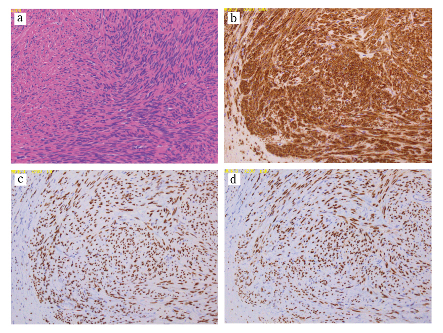
Although there have been a few theories, the exact pathophysiology of PM, other than iatrogenic factors, remains unclear. Hormonal stimulation is thought to be a key contributor in the pathophysiology of PM. Tavassoli et al also postulate that PM occurs due to an unusual predisposition of subperitoneal mesenchymal stem cells12). These processes are usually stimulated by estrogen12). This hypothesis is supported by cases of PM after the use of oral contraception13), hormone replacement therapy14), tamoxifen15), and ovarian stimulation treatment16). However, our patient received anti-hormonal therapy prior to undergoing TLH. As in our case, other reports describe the occurrence of PM without hormonal exposure, including a report of seven postmenopausal patients with PM, none of whom received hormone replacement therapy17). Based on these contradictory conclusions, the etiology of non-iatrogenic PM remains unclear. Although PM is usually a benign condition, nine cases of malignant transformation have been reported17). Among them, seven cases occurred in women of reproductive age17). Histopathological lesions, such as cytologic atypia, nuclear polymorphism, hyperchromasia, tumor cell necrosis, and increased mitotic Figures, are signs of malignant transformation17).


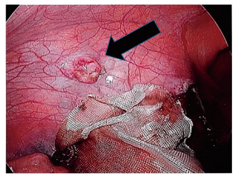
We perform laparoscopic surgery for an enlarged uterus in many cases, which allows for a detailed examination of various pelvic and abdominal conditions. Consequently, we found that PM could not be identified prior to performing surgery. We suggest that laparoscopic surgery enables the identification of small tumors growing in the peritoneal area.


In conclusion, we successfully managed uterine myoma and PM using laparoscopic surgery. Regardless of a history of previous myomectomy, PM should be considered in women with uterine myoma. This report also warrants further discussion and research into the biological characteristics of uterine myomas.

Conflict of interest disclosure

The authors have no potential conflicts of interest to declare.

Ethics

Informed consent was obtained from the patients for publication of this work.

Contributors

All authors were involved in the clinical care of the patient and contributed to the conception, drafting, review, and revision of the manuscript. All authors read and approved the final version of the paper and take full responsibility for the work.


Funding

This work did not receive any specific grant from funding agencies.

Provenance and peer review

This case report was peer reviewed.