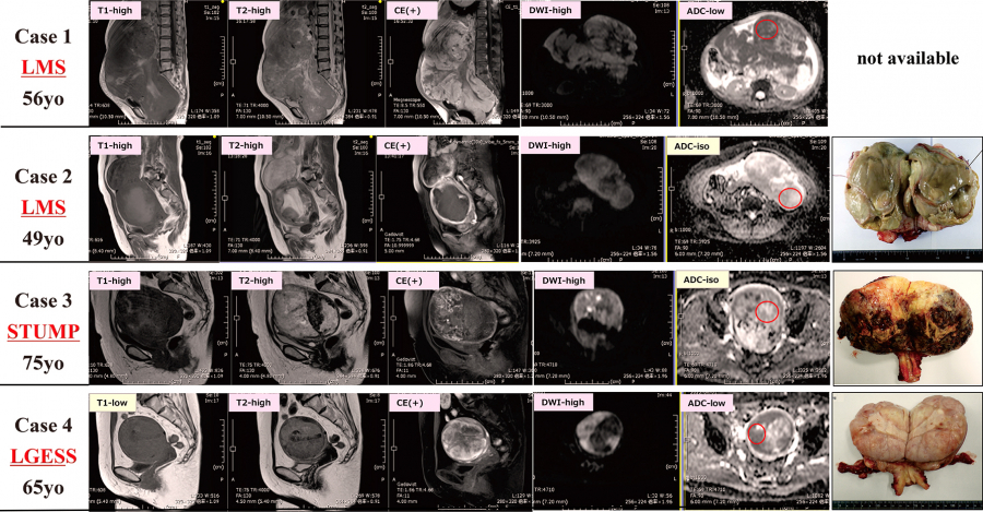
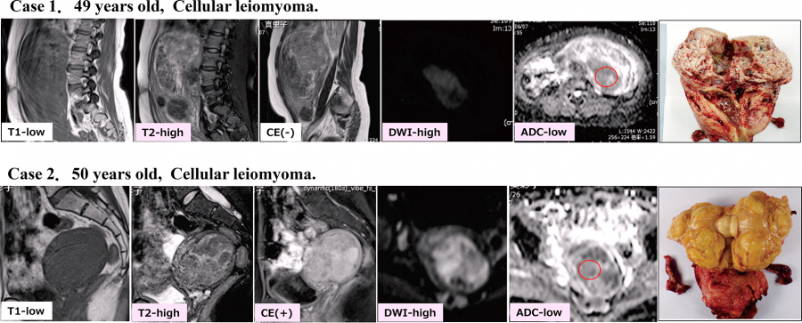
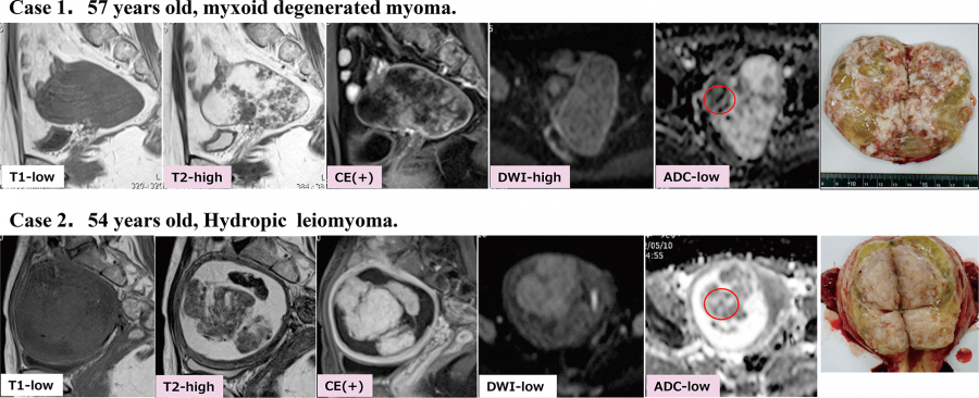
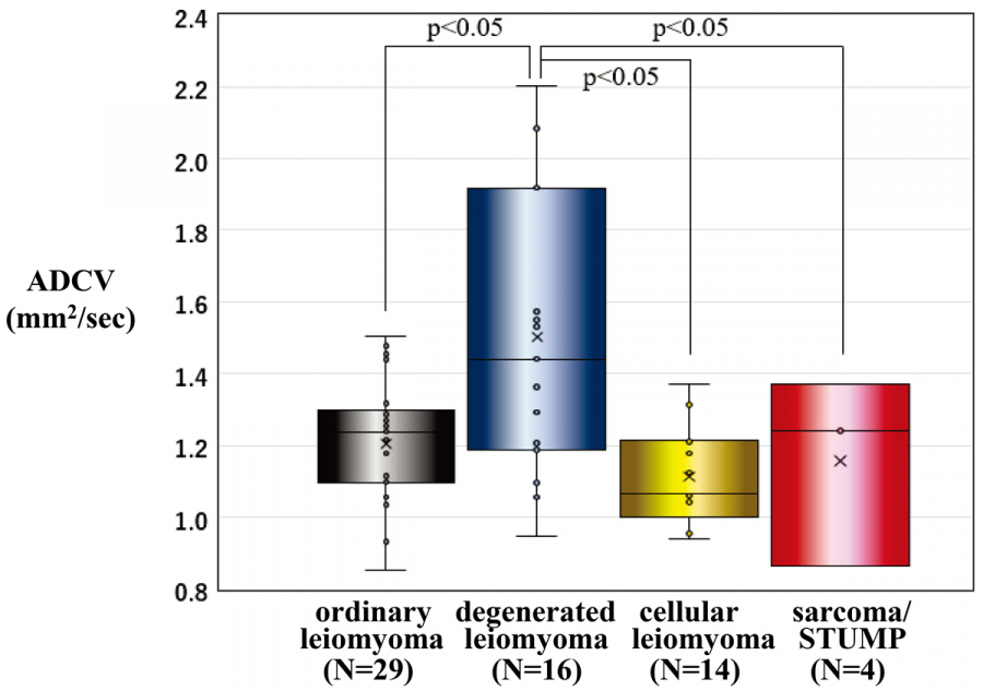
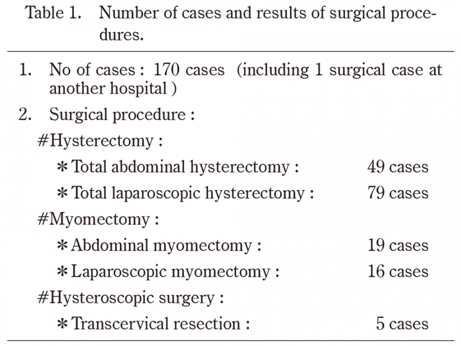
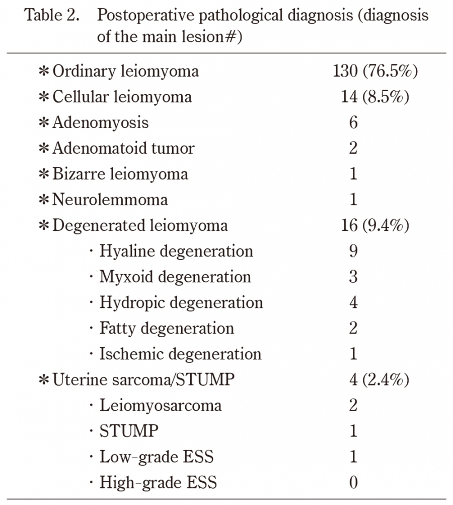
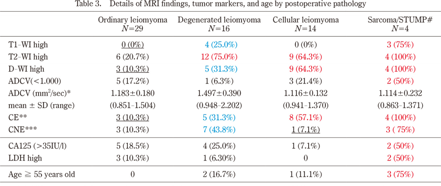
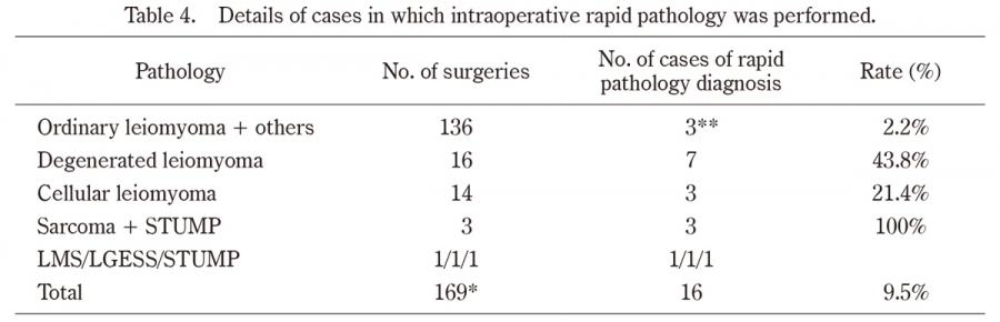
Although uterine sarcoma is a rare disease, its prognosis is extremely poor;thus, it is important to differentiate it from uterine leiomyoma. In this retrospective study, we examined the association between preoperative MRI findings and postoperative pathology results in 170 patients with uterine tumors who underwent preoperative MRI examination at Fukushima Red Cross Hospital. In 4 cases of sarcoma / smooth muscle tumor of unknown malignant potential (STUMP), abnormal findings were found at a high frequency with T1-weighted imaging (T1WI) (75%), T2-weighted imaging (T2WI), diffusion-weighted imaging (DWI), and contrast enhancement (CE) (100%). In cases of ordinary leiomyoma, on the other hand, abnormal findings were less frequent. The rates of high DWI signal intensity for degenerated and cellular leiomyoma were 31% and 64%, respectively, and the CE-positive rates were 31% and 57%, respectively. Apparent Diffusion Coefficient (ADC) values appeared to be useful in differentiating degenerated leiomyoma from sarcoma. The relatively characteristic findings of uterine sarcoma on MRI images may overlap with those of degenerated leiomyoma and cellular leiomyoma, making it difficult to diagnose sarcoma on imaging alone. However, findings that distinguish sarcoma from ordinary, degenerated, and cellular leiomyoma cases are worthy of attention, to avoid overlooking sarcoma.
Abstract/References
The utility of MRI for the preoperative differential diagnosis of uterine sarcoma and leiomyoma: a single-center study
Hiroyuki Yazawa, Riho Yazawa, Kazuki Anjo, Akari Inazuki, Manabu Kikuta
-
Hiroyuki Yazawa
Department of Obstetrics and Gynecology, Fukushima Red Cross Hospital
-
Riho Yazawa
Department of Obstetrics and Gynecology, Fukushima Red Cross Hospital
-
Kazuki Anjo
Junior Resident, Fukushima Red Cross Hospital
-
Akari Inazuki
Junior Resident, Fukushima Red Cross Hospital
-
Manabu Kikuta
Department of Radiology, Fukushima Red Cross Hospital
Abstract
References
1. Wang C, Zheng X, Zhou Z, Shi Y, Wu Q, Lin K. Differentiating cellular leiomyoma from uterine sarcoma and atypical leiomyoma using multi-parametric MRI. Front Oncol, 12:1005191, 2022. doi:10.3389/fonc.2022.1005191.
3. Marsh EE, Al-Hendy A, Kappus D, Galitsky A, Stewart EA. Kerolous MB, Prevalence, and Treatment of UFs:A survey of U.S.Women. J Women Health, 27:1359-1367, 2018.
5. Santos P, Cunha TM. Uterine sarcomas:clinical presentation and MRI features. Diagn Interv Radiol, 21:4-9, 2015. doi:https://doi.org/10.5152/dir.2014.14053.
6. Ando T, Kato H, Furui T, Morishige K, Goshima S, Matsuo M. Uterine smooth muscle tumors with hyperintense area on T1 weighted images: differentiation between leiomyosarcomas and leiomyomas. Br J Radiol, 91:20170767, 2018.
8. Li HM, Liu J, Qiang JW, Zhang H, Zhang GF, Ma F. Diffusion-weighted imaging for differentiating uterine leiomyosarcoma from degenerated leiomyoma. J Comput Assist Tomogr, 41:599-606, 2017.
10. Lin G, Yang LY, Huang YT, et al. Comparison of the diagnostic accuracy of contrast-enhanced MRI and diffusion-weighted MRI in the differentiation between uterine leiomyosarcoma/smooth muscle tumor with uncertain malignant potential and benign leiomyoma. J Magn Reson Imaging, 43, 333-342, 2015.
12. Wahab CA, Jannot AS, Bonaffini PA, Bourillon C, Cornou C, Lefrère-Belda MA, Bats AS, Thomassin-Naggara I, Belluci A, Reinhold C, Fournier LS. Diagnostic algorithm to differentiate benign atypical leiomyomas from malignant uterine sarcomas with diffusion-weighted MRI. Radiology, 297: 361-371, 2020.
Figures