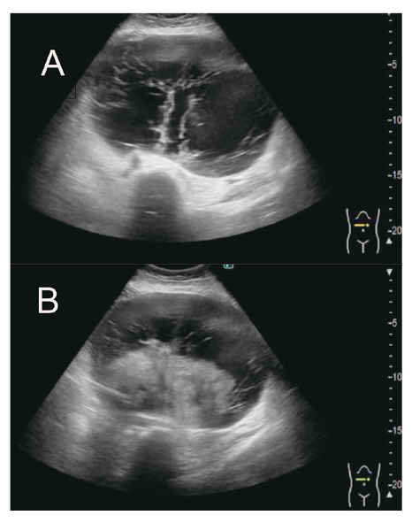
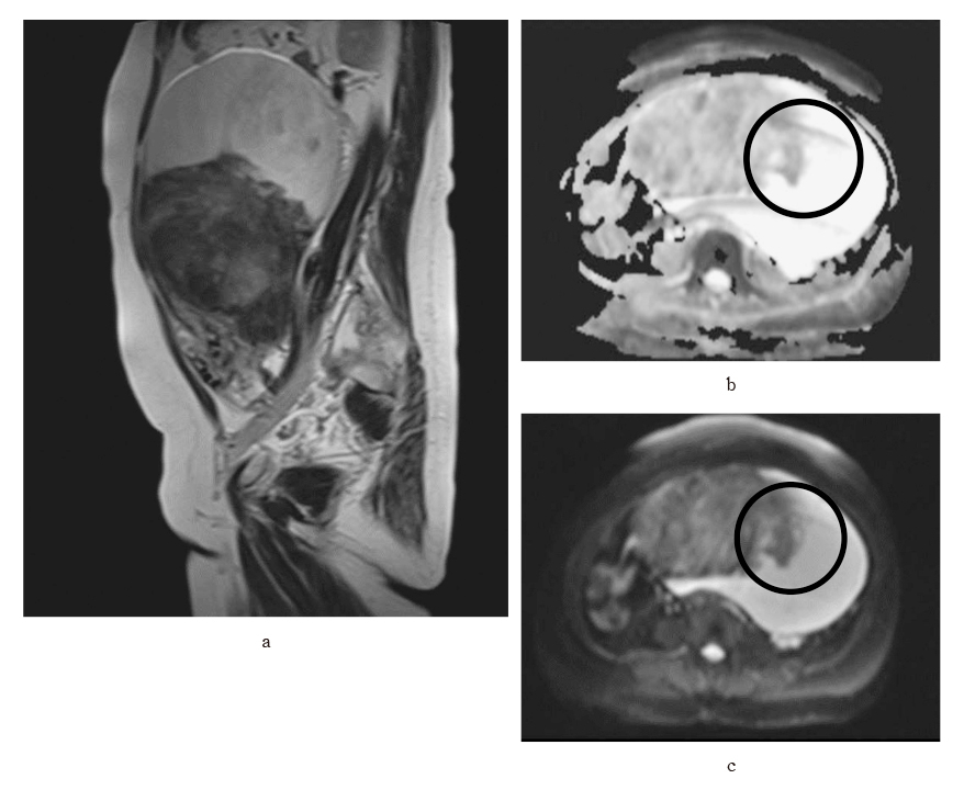
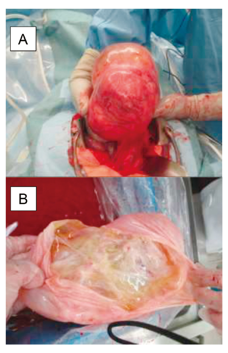
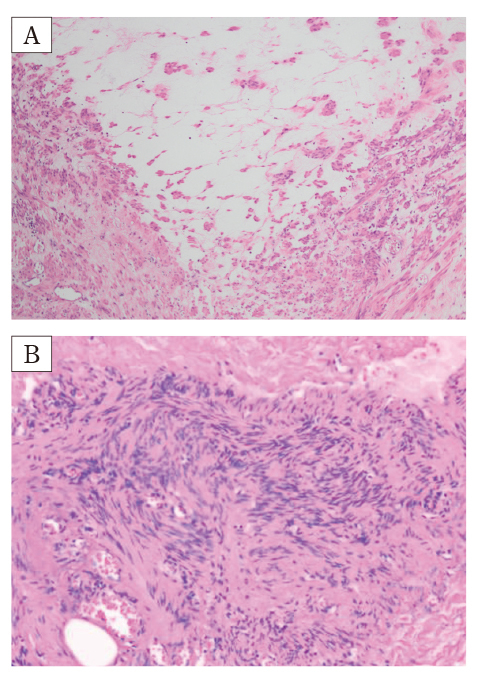
Uterine leiomyomas, benign tumors common in reproductive-aged women, can display rare variants such as hydropic leiomyoma (HL), which exhibit unique histological features like zonal edema and increased vascularity. However, due to its rarity, comprehensive clinical knowledge about HL is limited. We report a case of a 49-year-old Japanese woman who was premenopausal and nulliparous, presenting with a two-year history of abdominal distension. An MRI scan revealed a 20 cm mass in the posterior part of the uterus, exhibiting characteristics suggestive of an ovarian tumor. During laparotomy, a cystic tumor connected with a swollen fibroid was found, and pathology confirmed HL. This case emphasizes that hydropic leiomyomas can mimic malignant tumors on ultrasonography due to their atypical features, necessitating additional evaluations using alternative imaging techniques or histopathological examinations for accurate diagnosis and appropriate management. The patient recovered uneventfully, broadening our understanding of HL’s clinical presentation.
Abstract/References
Hydropic leiomyoma-like ovarian tumor: a case report
Tomoko Yamaguchi, Hyo Kyozuka, Yoshihiro Kochi, Fumihiro Ito, Hajime Odajima, Daisuke Suzuki, Yasuhisa Nomura
-
Tomoko Yamaguchi
Department of Obstetrics and Gynecology, Ohta Nisinouchi Hospital
-
Hyo Kyozuka
Department of Obstetrics and Gynecology, Ohta Nisinouchi Hospital
-
Yoshihiro Kochi
Department of Obstetrics and Gynecology, Ohta Nisinouchi Hospital
-
Fumihiro Ito
Department of Obstetrics and Gynecology, Ohta Nisinouchi Hospital
-
Hajime Odajima
Department of Pathology, Ohta Nisinouchi Hospital
-
Daisuke Suzuki
Department of Obstetrics and Gynecology, Ohta Nisinouchi Hospital
-
Yasuhisa Nomura
Department of Obstetrics and Gynecology, Ohta Nisinouchi Hospital
Abstract
References
2. D’Silva EC, Muda AM, Safiee AI, Ghazali WAHW. Five-Year Lapsed:Review of Laparoscopic Myomectomy versus Open Myomectomy in Putrajaya Hospital. Gynecol Minim Invasive Ther, 7:161-166, 2018.
3. Ghosh S, Naftalin J, Imrie R, Hoo WL. Natural History of Uterine Fibroids:A Radiological Perspective. Curr Obstet Gynecol Rep, 7:117-121, 2018.
4. Giannubilo SR, Ciavattini A, Petraglia F, Castellucci M, Ciarmela P. Management of fibroids in perimenopausal women. Curr Opin Obstet Gynecol, 27:416-421, 2015.
6. Babacan A, Kizilaslan C, Gun I, Muhcu M, Mungen E, Atay V. CA 125 and other tumor markers in uterine leiomyomas and their association with lesion characteristics. Int J Clin Exp Med, 7:1078-1083, 2014.
7. Sarkodie BD, Botwe BO, Ofori EK. Uterine fibroid characteristics and sonographic pattern among Ghanaian females undergoing pelvic ultrasound scan:a study at 3-major centres. BMC Womens Health, 16:10, 2016.
9. Horta M, Cunha TM, Oliveira R, Magro P. Hydropic leiomyoma of the uterus presenting as a giant abdominal mass. BMJ Case Rep, 2015:bcr2015211929, 2015.
11. Awad EE, El-agwany AS, Elhabashy AM, El-zarka A, Moneim ASA. A giant uterine myometrium cyst mimicking an ovarian cyst in pregnancy:An uncommon presentation of hydropic degeneration of uterine fibroid. The Egyptian Journal of Radiology and Nuclear Medicine, 46:529-534, 2015.
12. Moore L, Wilson S, Rosen B. Giant hydropic uterine leiomyoma in pregnancy:unusual sonographic and Doppler appearance. J Ultrasound Med, 13:416-418, 1994.
15. Roth LM, Reed RJ. Dissecting leiomyomas of the uterus other than cotyledonoid dissecting leiomyomas:a report of eight cases. Am J Surg Pathol, 23:1032-1039, 1999.
17. Abdel Wahab C, Jannot AS, Bonaffini PA, et al. Diagnostic algorithm to differentiate benign atypical leiomyomas from malignant uterine sarcomas with diffusion-weighted MRI. Radiology, 297:361-371, 2020.
19. Patil AR, Nandikoor S, Padilu R. Hydropic degeneration of leiomyoma in nongravid uterus:The “split fiber” sign on magnetic resonance imaging. Indian J Radiol Imaging, 28:182-186, 2018.
Figures