Abstract/References

Tau positron emission tomography in patients with cognitive impairment and suspected Alzheimer’s disease

Hiroshi Matsuda, Tensho Yamao

Author information
  • Hiroshi Matsuda

    Department of Biofunctional Imaging, Fukushima Medical University

  • Tensho Yamao

    Department of Radiological Sciences, School of Health Sciences, Fukushima Medical University

Abstract

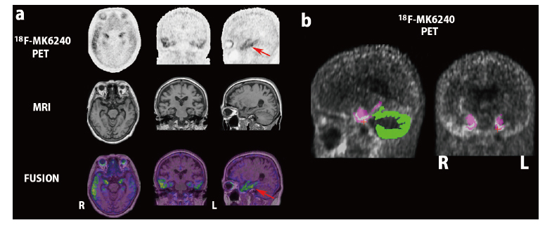
Alzheimer’s disease (AD) is diagnosed by the presence of both amyloid β and tau proteins. Recent advances in molecular PET imaging have made it possible to assess the accumulation of these proteins in the living brain. PET ligands have been developed that bind to 3R/4R tau in AD, but not to 3R tau or 4R tau alone. Of the first-generation PET ligands, 18F-flortaucipir has recently been approved by the Food and Drug Administration. Several second-generation PET probes with less off-target binding have been developed and are being applied clinically. Visual interpretation of tau PET should be based on neuropathological neurofibrillary tangle staging instead of a simple positive or negative classification. Four visual read classifications have been proposed: “no uptake,” “medial temporal lobe (MTL) only,” “MTL AND,” and “outside MTL.” As an adjunct to visual interpretation, quantitative analysis has been proposed using MRI-based native space FreeSurfer parcellations. The standardized uptake value ratio of the target area is measured using the cerebellar gray matter as a reference region. In the near future, the Centiloid scale of tau PET is expected to be used as a harmonized value for standardizing each analytical method or PET ligand used, similar to amyloid PET.

The cintent of reseach paper

References

1. Jack CR Jr, Bennett DA, Blennow K, et al. Toward a biological definition of Alzheimer’s disease. Alzheimers Dement, 14:535-562, 2018.


2. Nelson PT, Head E, Schmitt FA, et al. Alzheimer’s disease is not “brain aging”: neuropathological, genetic, and epidemiological human studies. Acta Neuropathol, 121:571-587, 2011.


3. Serrano-Pozo A, Qian J, Monsell SE, et al. Mild to moderate Alzheimer dementia with insufficient neuropathological changes. Ann Neurol, 75:597-601, 2014.


4. Nelson PT, Alafuzoff I, Bigio EH, et al. Correlation of Alzheimer disease neuropathologic changes with cognitive status:a review of the literature. J Neuropathol Exp Neurol, 71:362-381, 2012.


5. Jack CR Jr, Bennett DA, Blennow K, et al. A/T/N:An unbiased descriptive classification scheme for Alzheimer disease biomarkers. Neurology, 87:539-547, 2016.


6. Matsuda H, Shigemoto Y, Sato N. Neuroimaging of Alzheimer’s disease:focus on amyloid and tau PET. Jpn J Radiol, 37:735-749, 2019.


7. Rabinovici GD, Gatsonis C, Apgar C, et al. Association of amyloid positron emission tomography with subsequent change in clinical management among Medicare beneficiaries with mild cognitive impairment or dementia. JAMA, 321:1286-1294, 2019.


8. Matsuda H, Ito K, Ishii K, et al. Quantitative evaluation of 18F-Flutemetamol PET in patients with cognitive impairment and suspected Alzheimer’s disease:a multicenter study. Front Neurol, 11:578753, 2021.


9. Matsuda H, Okita K, Motoi Y, et al. Clinical impact of amyloid PET using 18F-florbetapir in patients with cognitive impairment and suspected Alzheimer’s disease:a multicenter study. Ann Nucl Med, 36:1039-1049, 2022


10. Beyer L, Brendel M. Imaging of tau pathology in neurodegenerative diseases:an update. Semin Nucl Med, 51:253-263, 2021.


11. Cassinelli Petersen G, Roytman M, Chiang GC, Li Y, Gordon ML, Franceschi AM. Overview of tau PET molecular imaging. Curr Opin Neurol, 35:230-239, 2022.


12. Wang Y, Mandelkow E. Tau in physiology and pathology. Nat Rev Neurosci, 17:5-21, 2016.


13. Zhang Y, Wu KM, Yang L, et al. Tauopathies:new perspectives and challenges. Mol Neurodegeneration, 17:28, 2022.


14. Shoghi-Jadid K, Small GW, Agdeppa ED, et al. Localization of neurofibrillary tangles and beta-amyloid plaques in the brains of living patients with Alzheimer disease. Am J Geriatr Psychiatry, 10:24-35, 2002.


15. Maruyama M, Shimada H, Suhara T, et al. Imaging of tau pathology in a tauopathy mouse model and in Alzheimer patients compared to normal controls. Neuron, 79:1094-1108, 2013.


16. Hashimoto H, Kawamura K, Takei M, et al. Identification of a major radiometabolite of [11C]PBB3. Nucl Med Biol, 42:905-910, 2015.


17. Okamura N, Furumoto S, Harada R, et al. Novel 18F-labeled arylquinoline derivatives for noninvasive imaging of tau pathology in Alzheimer disease. J Nucl Med, 54:1420-1427, 2013.


18. Harada R, Okamura N, Furumoto S, et al. 18F-THK5351:A Novel PET Radiotracer for Imaging Neurofibrillary Pathology in Alzheimer Disease. J Nucl Med, 57:208-214, 2016.


19. Ng KP, Pascoal TA, Mathotaarachchi S, et al. Monoamine oxidase B inhibitor, selegiline, reduces 18F-THK5351 uptake in the human brain. Alzheimers Res Ther, 9:25, 2017.


20. Marquié M, Normandin MD, Vanderburg CR, et al. Validating novel tau positron emission tomography tracer [F-18]-AV-1451 (T807) on postmortem brain tissue. Ann Neurol, 78:787-800, 2015.


21. Mattay VS, Fotenos AF, Ganley CJ, Marzella L. Brain tau Imaging:Food and Drug Administration approval of 18F-Flortaucipir Injection. J Nucl Med, 61:1411-1412, 2020.


22. Jie CVML, Treyer V, Schibli R, Mu L. Tauvid™: The first FDA-approved PET tracer for imaging tau pathology in Alzheimer’s disease. Pharmaceuticals (Basel), 14:110, 2021.


23. Sander K, Lashley T, Gami P, et al. Characterization of tau positron emission tomography tracer [18F]AV-1451 binding to postmortem tissue in Alzheimer’s disease, primary tauopathies, and other dementias. Alzheimers Dement, 12:1116-1124, 2016.


24. Lowe VJ, Curran G, Fang P, et al. An autoradiographic evaluation of AV-1451 Tau PET in dementia. Acta Neuropathol Commun, 4:58, 2016.


25. Marquié M, Verwer EE, Meltzer AC, et al. Lessons learned about [F-18]-AV-1451 off-target binding from an autopsy-confirmed Parkinson’s case. Acta Neuropathol Commun, 5:75, 2017.


26. Lee CM, Jacobs HIL, Marquié M, et al. 18F-Flortaucipir Binding in Choroid Plexus:Related to Race and Hippocampus Signal. J Alzheimers Dis, 62:1691-1702, 2018.


27. Ikonomovic MD, Uryu K, Abrahamson EE, et al. Alzheimer’s pathology in human temporal cortex surgically excised after severe brain injury. Exp Neurol, 190:192-203, 2004.


28. Choi JY, Cho H, Ahn SJ, et al. Off-Target 18F-AV-1451 Binding in the Basal Ganglia Correlates with Age-Related Iron Accumulation. J Nucl Med, 59:117-120, 2018.


29. Ikonomovic MD, Abrahamson EE, Price JC, Mathis CA, Klunk WE. [F-18]AV-1451 positron emission tomography retention in choroid plexus: More than “off-target” binding. Ann Neurol, 80:307-308, 2016.


30. Vermeiren C, Motte P, Viot D, Mairet-Coello G, et al. The tau positron-emission tomography tracer AV-1451 binds with similar affinities to tau fibrils and monoamine oxidases. Mov Disord, 33:273-281, 2018.


31. Hansen AK, Brooks DJ, Borghammer P. MAO-B Inhibitors do not block in vivo Flortaucipir([18F]-AV-1451) binding. Mol Imaging Biol, 20:356-360, 2018.


32. Yap SY, Frias B, Wren MC, et al. Discriminatory ability of next-generation tau PET tracers for Alzheimer’s disease. Brain, 144:2284-2290, 2021.


33. Honer M, Gobbi L, Knust H, et al. Preclinical Evaluation of 18F-RO6958948, 11C-RO6931643, and 11C-RO6924963 as Novel PET Radiotracers for Imaging Tau Aggregates in Alzheimer Disease. J Nucl Med, 59:675-681, 2018.


34. Wong DF, Comley RA, Kuwabara H, et al. Characterization of 3 Novel Tau Radiopharmaceuticals, 11C-RO-963, 11C-RO-643, and 18F-RO-948, in Healthy Controls and in Alzheimer Subjects. J Nucl Med, 59:1869-1876, 2018.


35. Kuwabara H, Comley RA, Borroni E, et al. Evaluation of 18F-RO-948 PET for Quantitative Assessment of Tau Accumulation in the Human Brain. J Nucl Med, 59:1877-1884, 2018.


36. Sanabria Bohórquez S, Marik J, Ogasawara A, et al. [18F]GTP1 (Genentech Tau Probe 1), a radioligand for detecting neurofibrillary tangle tau pathology in Alzheimer’s disease. Eur J Nucl Med Mol Imaging, 46:2077-2089, 2019.


37. Blennow K, Chen C, Cicognola C, et al. Cerebrospinal fluid tau fragment correlates with tau PET:a candidate biomarker for tangle pathology. Brain, 143:650-660, 2020.


38. Kroth H, Oden F, Molette J, et al. Discovery and preclinical characterization of [18F]PI-2620, a next-generation tau PET tracer for the assessment of tau pathology in Alzheimer’s disease and other tauopathies. Eur J Nucl Med Mol Imaging, 46:2178-2189, 2019.


39. Mormino EC, Toueg TN, Azevedo C, et al. Tau PET imaging with 18F-PI-2620 in aging and neurodegenerative diseases. Eur J Nucl Med Mol Imaging, 48:2233-2244, 2021.


40. Baker SL, Provost K, Thomas W, et al. Evaluation of [18F]-JNJ-64326067-AAA tau PET tracer in humans. J Cereb Blood Flow Metab, 41:3302-3313, 2021.


41. Tagai K, Ono M, Kubota M, et al. High-Contrast In Vivo Imaging of Tau Pathologies in Alzheimer’s and Non-Alzheimer’s Disease Tauopathies. Neuron, 109:42-58.e8, 2021.


42. Ishizuchi K, Takizawa T, Tezuka T, et al. A case of progressive supranuclear palsy with predominant cerebellar ataxia diagnosed by [18F]PM-PBB3 tau PET. J Neurol Sci, 425:117440, 2021.


43. Zhou Y, Li J, Nordberg A, Ågren H. Dissecting the binding profile of PET tracers to corticobasal degeneration tau fibrils. ACS Chem Neurosci, 12:3487-3496, 2021.


44. Mishra SK, Yamaguchi Y, Higuchi M, Sahara N. Pick’s tau fibril shows multiple distinct PET probe binding sites:insights from computational modelling. Int J Mol Sci, 22:349, 2020.


45. Knight AC, Morrone CD, Varlow C, Yu WH, McQuade P, Vasdev N. Head-to-head comparison of tau-PET radioligands for imaging TDP-43 in post-mortem ALS brain. Mol Imaging Biol, 2022;10.1007/s11307-022-01779-1.


46. Perez-Soriano A, Arena JE, Dinelle K, et al. PBB3 imaging in Parkinsonian disorders:Evidence for binding to tau and other proteins. Mov Disord, 32:1016-1024, 2017.


47. Koga S, Ono M, Sahara N, Higuchi M, Dickson DW. Fluorescence and autoradiographic evaluation of tau PET ligand PBB3 to α-synuclein pathology. Mov Disord, 32:884-892, 2017.


48. Walji AM, Hostetler ED, Selnick H, et al. Discovery of 6-(Fluoro-(18)F)-3-(1H-pyrrolo[2,3-c]pyridin-1-yl)isoquinolin-5-amine ([(18)F]-MK-6240):A Positron Emission Tomography (PET) Imaging Agent for Quantification of Neurofibrillary Tangles (NFTs). J Med Chem, 59:4778-4789, 2016.


49. Leuzy A, Pascoal TA, Strandberg O, et al. A multicenter comparison of [18F]flortaucipir, [18F]RO948, and [18F]MK6240 tau PET tracers to detect a common target ROI for differential diagnosis. Eur J Nucl Med Mol Imaging, 48:2295-2305, 2021.


50. Hostetler ED, Walji AM, Zeng Z, et al. Preclinical characterization of 18F-MK-6240, a promising PET tracer for In vivo quantification of human neurofibrillary tangles. J Nucl Med, 57:1599-1606, 2016.


51. Pascoal TA, Shin M, Kang MS, et al. In vivo quantification of neurofibrillary tangles with [18F]MK-6240. Alzheimers Res Ther, 10:74, 2018.


52. Betthauser TJ, Cody KA, Zammit MD, et al. In vivo characterization and quantification of neurofibrillary tau PET radioligand 18F-MK-6240 in humans from Alzheimer disease dementia to young controls. J Nucl Med, 60:93-99, 2019.


53. Lohith TG, Bennacef I, Vandenberghe R, et al. Brain imaging of Alzheimer dementia patients and elderly controls with 18F-MK-6240, a PET tracer targeting neurofibrillary tangles. J Nucl Med, 60:107-114, 2019.


54. Pascoal TA, Therriault J, Benedet AL, et al. 18F-MK-6240 PET for early and late detection of neurofibrillary tangles. Brain, 143:2818-2830, 2020.


55. Aguero C, Dhaynaut M, Normandin MD, et al. Autoradiography validation of novel tau PET tracer [F-18]-MK-6240 on human postmortem brain tissue. Acta Neuropathol Commun, 7:37, 2019.


56. Salinas C, Lohith TG, Purohit A, et al. Test-retest characteristic of [18F]MK-6240 quantitative outcomes in cognitively normal adults and subjects with Alzheimer’s disease. J Cereb Blood Flow Metab, 40:2179-2187, 2020.


57. Braak H, Braak E. Neuropathological stageing of Alzheimer-related changes. Acta Neuropathol, 82:239-259, 1991.


58. Braak H, Alafuzoff I, Arzberger T, Kretzschmar H, Del Tredici K. Staging of Alzheimer disease-associated neurofibrillary pathology using paraffin sections and immunocytochemistry. Acta Neuropathol, 112:389-404, 2006.


59. Gogola A, Minhas DS, Villemagne VL, et al. Direct Comparison of the Tau PET Tracers 18F-Flortaucipir and 18F-MK-6240 in Human Subjects. J Nucl Med, 63:108-116, 2022.


60. Shuping JL, Matthews DC, Adamczuk K, et al. Development, initial validation, and application of a visual read method for [18F]MK-6240 tau PET. Alzheimers Dement (NY). 9:e12372, 2023.


61. Bennett RE, DeVos SL, Dujardin S, et al. Enhanced Tau Aggregation in the Presence of Amyloid β. Am J Pathol, 187:1601-1612, 2017.


62. Vogel JW, Iturria-Medina Y, Strandberg OT, et al. Spread of pathological tau proteins through communicating neurons in human Alzheimer’s disease. Nat Commun, 11:2612, 2020.


63. Crary JF, Trojanowski JQ, Schneider JA, et al. Primary age-related tauopathy (PART):a common pathology associated with human aging. Acta Neuropathol, 128:755-766, 2014.


64. Krishnadas N, Doré V, Groot C, et al. Mesial temporal tau in amyloid-β-negative cognitively normal older persons. Alzheimers Res Ther, 14:51, 2022.


65. Krishnadas N, Huang K, Schultz SA, et al. Visually Identified Tau 18F-MK6240 PET Patterns in Symptomatic Alzheimer’s Disease. J Alzheimers Dis, 88:1627-1637, 2022.


66. Villemagne VL, Doré V, Bourgeat P, et al. The Tau MeTeR composites for the generation of continuous and categorical measures of tau deposits in the brain. J Mol Med Ther, 1:25-32, 2017.


67. Harrison TM, Ward TJ, Murphy A, et al. Optimizing quantification of MK6240 tau PET in unimpaired older adults. Neuroimage, 265:119761, 2023.


68. Yamao T, Miwa K, Wagatsuma K, et al. Centiloid scale analysis for 18F-THK5351 PET imaging in Alzheimer’s disease. Physica medica, 82:249-254, 2021.


69. Mintun MA, Lo AC, Duggan Evans C, et al. Donanemab in Early Alzheimer’s Disease. N Engl J Med, 384:1691-1704, 2021.


70. Rafii MS, Sperling RA, Donohue MC, et al. The AHEAD 3-45 Study:Design of a prevention trial for Alzheimer’s disease. Alzheimers Dement, 19:1227-1233, 2023.

Figures