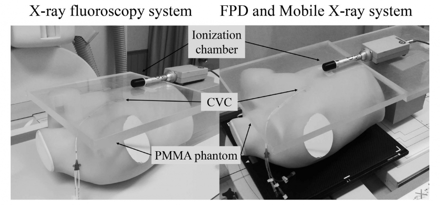
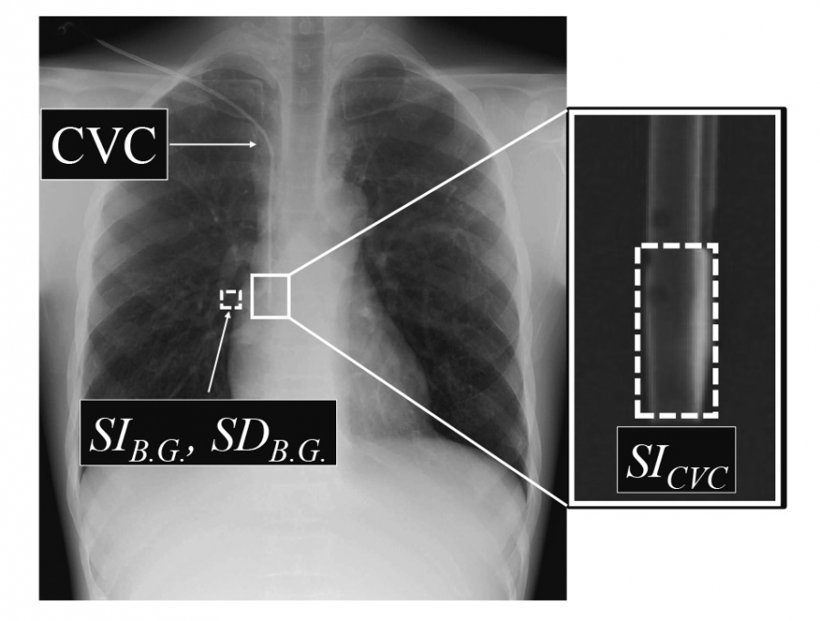
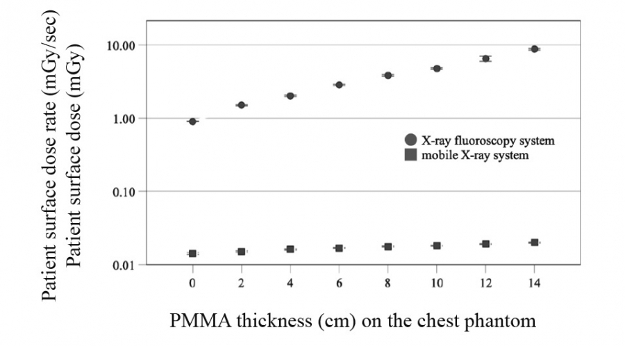
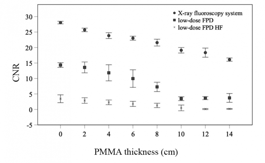
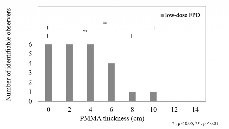
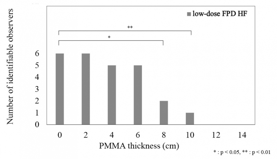
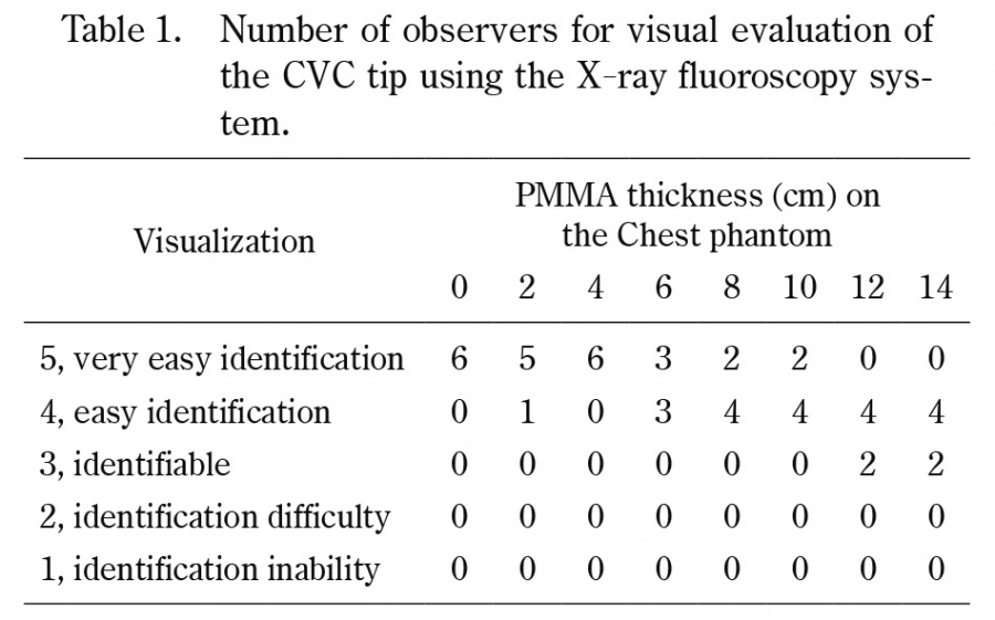
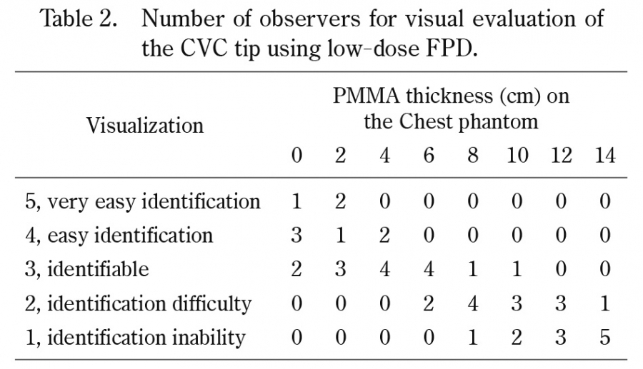
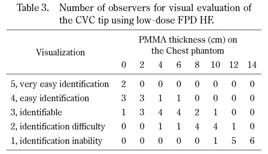
A central venous catheter (CVC) should be inserted at the optimum position to infuse medicines, blood products, nutrients, or fluids. Positioning of the catheter tip is commonly performed under landmark or fluoroscopic guidance. However, Japanese regulations do not allow the performing of fluoroscopy-guided procedures outside of the fluoroscopy room. We hypothesized that a new image-guided CVC placement technique by combining a wireless flat-panel detector (FPD) and a mobile X-ray system could be applied at the bedside to support CVC insertion. A CVC attached to a chest phantom in conjunction with the polymethyl methacrylate (PMMA) phantom was imaged, contrast-to-noise ratio (CNR) was measured with images, and radiologists and emergency physicians rated the catheter images using a Likert scale for visual evaluation. The minimum dose of the FPD and mobile X-ray system was reduced by at least 98% compared with that of the X-ray fluoroscopy system. The CNR decreased with the increasing PMMA phantom thickness. However, results of the visual evaluation were maintained at the clinically usable score with low-dose imaging up to a 6-cm thickness of the PMMA phantom. In conclusion, the combination of FPD and mobile X-ray systems is particularly effective in the emergency room setting where such procedures are required to be performed with urgency.
Abstract/References
A proposed combination of flat-panel detector and mobile X-ray systems for low-dose image-guided central venous catheter insertion
Masami Tashiro, Hitoshi Kubo, Chie Kanezawa, Hiroshi Ito
-
Masami Tashiro
Department of Radiological Sciences, School of Health Science, Fukushima Medical University
Department of Radiology and Nuclear Medicine, Fukushima Medical University -
Hitoshi Kubo
Department of Radiological Sciences, School of Health Science, Fukushima Medical University
-
Chie Kanezawa
Department of Radiology, Fukushima Medical University
-
Hiroshi Ito
Department of Radiology and Nuclear Medicine, Fukushima Medical University
Abstract
References
1. Pires R, Rodrigues N, Machado J, Cruz R. Central venous catheterization:an updated review of historical aspects, indications, techniques, and complications. Transl Surgery, 2:66-70, 2017.
2. Dudrick SJ, Wilmore DW, Vars HM, Rhoads JE. Long-term total parenteral nutrition with growth, development, and positive nitrogen balance. Surgery, 64:134-142, 1968.
3. Filler RM, Eraklis AJ, Rubin VG, Das GB. Long-term total parenteral nutrition in infants. N Engl J Med, 281:589-594, 1969.
4. Padberg FT Jr, Ruggiero J, Blackburn GL, Bistrian BR. Central venous catheterization for parenteral nutrition. Annals Surg, 193:264-270, 1981.
5. Fukuda S, Nakajima K, Miyazaki Y, et al. Use of double-lumen peripherally inserted central catheters for safer perioperative management of esophageal cancer patients. J Vasc Access, 16:338-343, 2015.
6. Fang S, Yang J, Song L, et al. Comparison of three types of central venous catheters in patients with malignant tumor receiving chemotherapy. Patient Prefer Adherence, 11:1197-1204, 2017.
8. Toshniwal GR, Rath GP, Bithal PK. Flush test—a new technique to assess the malposition of subclavian central venous catheter position in the internal jugular vein. J Neurosurg Anesthesiol, 18:268-269, 2006.
9. Kuchle C, Suttmann Y, Wen M. Placement of central venous dialysis catheters without X-ray: safety and feasibility. J Nephrol Ren Dis, 1, 2017.
13. Watabe O, Kimura T, Okada K, et al. The safety and reliability of central venous catheterization by means of axillary vein puncture with real-time ultrasound and fluoroscopic guidance. J Jpn Soc Intensive Care Med, 16:163-167, 2009.
15. Baba M, Goto M, Hirano M, et al. Investigation of image processing using “catheter/gauze enhancement function” (Japanese). Saitama Housyasen, 62:397-404, 2014.
18. Marasco SF, Lukas G, McDonald M, et al. Review of ECMO (extra corporeal membrane oxygenation) support in critically ill adult patients. Heart Lung Circ, 17:S41-S47, 2008.
Figures (11)