Abstract/References

Different prognostic outcomes in two cases of FDG-PET/CT-Positive and -negative cardiac angiosarcoma

Ryo Yamakuni, Shiro Ishii, Shoki Yamada, Junko Hara, Hiroki Suenaga, Shigeyasu Sugawara, Hirofumi Sekino, Takayoshi Yamaki, Keiichi Ishida, Yuko Hashimoto, Hiroshi Ito

Author information
  • Ryo Yamakuni

    Department of Radiology and Nuclear Medicine, Fukushima Medical University

  • Shiro Ishii

    Department of Radiology and Nuclear Medicine, Fukushima Medical University

  • Shoki Yamada

    Department of Diagnostic Pathology, Fukushima Medical University

  • Junko Hara

    Department of Radiology and Nuclear Medicine, Fukushima Medical University

  • Hiroki Suenaga

    Department of Radiology and Nuclear Medicine, Fukushima Medical University

  • Shigeyasu Sugawara

    Department of Radiology and Nuclear Medicine, Fukushima Medical University

  • Hirofumi Sekino

    Department of Radiology and Nuclear Medicine, Fukushima Medical University

  • Takayoshi Yamaki

    Department of Cardiovascular Medicine, Fukushima Medical University

  • Keiichi Ishida

    Department of Cardiovascular Surgery, Fukushima Medical University

  • Yuko Hashimoto

    Department of Diagnostic Pathology, Fukushima Medical University

  • Hiroshi Ito

    Department of Radiology and Nuclear Medicine, Fukushima Medical University

Abstract

Cardiac angiosarcoma is a rare malignant tumor with a poor prognosis, characterized by the high uptake of 18F-fluorodeoxyglucose (FDG). This case report presents two cases of cardiac angiosarcoma with a marked difference in FDG uptake and prognosis.


Case Summary:


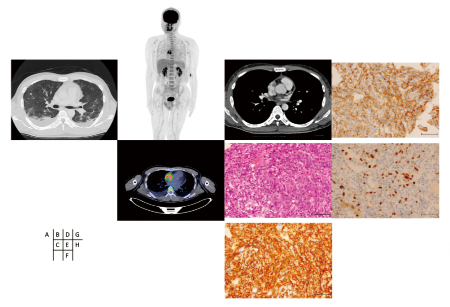
Case 1: A 40-year-old male presented with syncope. Ultrasound echocardiography demonstrated a cardiac tumor with a high uptake of 18F-FDG (maximum standardized uptake value=9.2). The patient underwent heart catheterization and tumor biopsy. The pathological result was high-grade angiosarcoma, and the MIB-1(Ki-67) proliferation index was approximately 20%. Systemic chemotherapy was administered; however, the patient died 2 years and 5 months after disease onset.


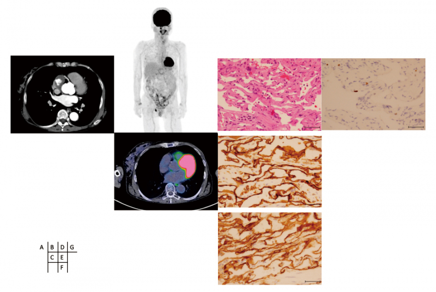
Case 2: A 65-year-old female had a right atrial tumor incidentally diagnosed during routine ultrasound echocardiography. The tumor exhibited a low uptake of 18F-FDG (maximum standardized uptake value=1.8). Open heart surgery was performed, and the tumor was completely resected. Histological analysis revealed low-grade angiosarcoma, and the MIB-1(Ki-67) proliferation index was less than 5%. The patient was followed-up and had not relapsed 2 years after surgery.


Conclusion: 18F-FDG uptake may reflect pathological tumor grade and prognosis in cardiac angiosarcoma.

The cintent of reseach paper

References

1. Silverman NA. Primary cardiac tumors. Ann Surg, 191(2):127-138, 1980.


2. McAllister HA. Primary tumors of the heart and pericardium. Pathol Annu, 14(2):325-355, 1979.


3. Aboud A, Farha K, Hsieh WC, et al. Prognostic factors for long-term survival after surgical resection of primary cardiac sarcoma. Thorac Cardiovasc Surg, 67(8):665-671, 2019.


4. Patel SD, Peterson A, Bartczak A, et al. Primary cardiac angiosarcoma - a review. Med Sci Monit, 20:103-109, 2014.


5. Shanmugam G. Primary cardiac sarcoma. Eur J Cardiothorac Surg, 29(6):925-932, 2006.


6. Misaki T, Ishizu K, Ishimori T, Takashi K. Gamut of FDG-PET. Japanese J Nucl Med, 49(4):357-389, 2012.


7. Ordóñez NG. Immunohistochemical endothelial markers:A review. Adv Anat Pathol, 19(5):281-295, 2012.


8. Spyratos F, Ferrero-Poüs M, Trassard M, et al. Correlation between MIB-1 and other proliferation markers:Clinical implications of the MIB-1 cutoff value. Cancer, 94(8):2151-2159, 2002.


9. Pauwels EKJ, Ribeiro MJ, Stoot JHMB, McCready VR, Bourguignon M, Mazière B. FDG accumulation and tumor biology. Nucl Med Biol, 25(4):317-322, 1998.


10. Rahbar K, Seifarth H, Schäfers M, et al. Differentiation of malignant and benign cardiac tumors using 18 F-FDG PET/CT. J Nucl Med, 53(6):856-863, 2012.


11. Kato A, Nakamoto Y, Ishimori T, Saga T, Togashi K. Prognostic value of quantitative parameters of 18F-FDG PET/CT for patients with angiosarcoma. AJR Am J Roentgenol, 214(3):649-657, 2020.


12. Nguyen XC, Lee WW, Chung JH, et al. FDG uptake, glucose transporter type 1, and Ki-67 expressions in non-small-cell lung cancer:correlations and prognostic values. Eur J Radiol, 62(2):214-219, 2007.


13. Bromińska B, Czepczyński R, Gabryel P, et al. 18F-FDG PET/CT and nestin expression as prognostic tools in pulmonary neuroendocrine tumours. Nucl Med Commun, 40(4):353-360, 2019.

Figures